


Support during Immunotherapy
Contents:
- What is support during immunotherapy
- Clinical decision support and pharmacokinetics
- Practical suggestions for support during immunotherapy
- Immune-related side effects
- Biological half-lives – Links
Summary of Support during Immunotherapy
The microbiome as the engine:
- A healthy gut flora, including the bacterium Akkermansia, is crucial for immunotherapy (checkpoint inhibitors) to activate the immune system against cancer cells at all.
Managing side effects:
- By identifying early signs of immune-related inflammation in the gut, lungs and skin, you can support the body’s tolerance without weakening the treatment effect.
Selective protection:
- With combination treatment (chemo + immunotherapy), the support strategy must protect the organs from chemo toxicity while preserving the immune system’s drive to attack.
Synergy through balance:
- Targeted use of prebiotics and specific antioxidants can optimise the therapeutic window and reduce the risk of treatment interruptions.
What is support during immunotherapy
Immunotherapy has revolutionised oncology by not attacking the cancer directly, but by releasing the “brake” on the body’s own immune system. This mechanism requires a specific environment to function optimally. The immune system needs “intelligence” from the gut to work. A healthy microbiome sends chemical signals that activate T cells and teach them to recognise cancer cells. Without these bacteria, immunotherapy is like pressing the accelerator in a car without fuel—the medicine releases the brake (PD-1), but the immune system has neither the energy nor the instruction to attack. [118].
The challenge is twofold
First, not all patients respond to treatment, which is often due to deficiencies in the gut flora. Second, the activated immune system can become overactive and attack healthy organs—a condition known as immune-related adverse events [119].
See also The Microbiome and Diet
Mechanisms of immune support

The microbiome axis
Research shows that the presence of specific bacteria such as Akkermansia muciniphila acts as a catalyst for treatment. Without these bacteria, the immune system may struggle to recognise the tumour [130].
Reducing side effects: irAEs (immune toxicity)
When the immune system attacks healthy tissue, inflammation occurs. The strategy here is to use specific substances that support tissue integrity (e.g., in the gut or skin) without suppressing the entire immune response [131].
Combination synergy
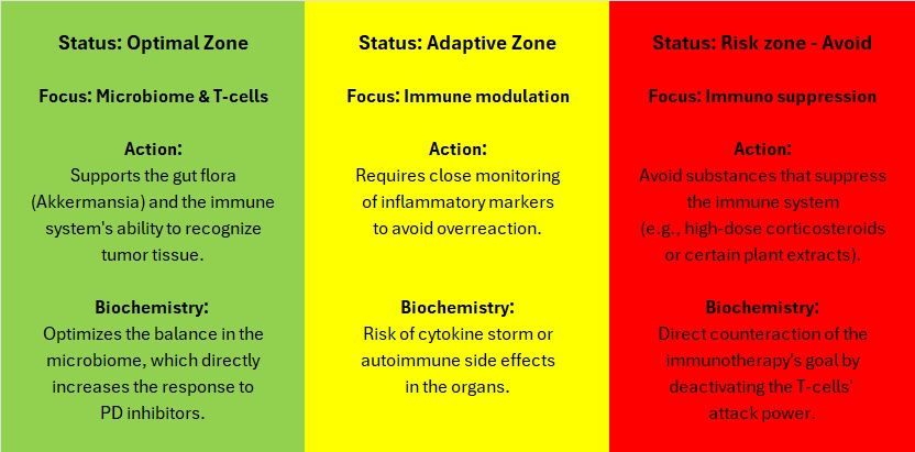
When immunotherapy is given together with chemotherapy, support must be balanced. You must protect the organs from chemo toxicity (as described in the article on Organ Protection), but avoid high-dose antioxidants or anti-inflammatory agents within the “checkpoint window”, as this can deactivate immune cells [28].
Safety zones for immunotherapy
See also Quality of life and shared responsibility
See also Safe measures
❤
What you read on I Have Cancer is not a recommendation. Seek qualified guidance.
How to use the article
This article is based on the principles of safety distance (washout) and interaction risk described in Chemo- and Radiotherapy Support. The purpose is to ensure that the supplementary measures support blood formation without interfering with the oncological treatment. Two tools are used to ensure full treatment integrity:
- Risk graduation
- Half-life
First and foremost, one must look at the interaction risk. The higher this is, the more importance should be attributed to the half-life. That is to say, if there is no risk of interaction, the half-life is of less importance (however, the substance must still be broken down and excreted, which can burden the organism). And if there is a high risk of interaction, the half-life becomes extremely crucial.
Risk assessment
Risk of interaction
The risk is assessed for both Chemotherapy (impact on medication and liver) and Radiotherapy (impact on the sensitivity of tumor cells).
Grading:
- None: The preparation does not interfere with the treatment.
- Low: Small risk, which is eliminated by following the safety distance.
- Moderate: Clear biological effect. Pauses must be strictly observed.
Biological half-life
The color codes illustrate the pause based on how quickly the substance is broken down and excreted. [14, 27]:
- Green: ◯ Fast out of the body (under 4 hours). High degree of control.
- Yellow: ⬤ Longer time to be broken down/excreted (4–24 hours). Requires a pause of 1–5 days.
Acute vs. accumulated dose
Be aware that half-lives are often based on a single dose. With regular use of certain supplements (e.g., fat-soluble vitamins), the substance can accumulate in the tissues, which may require a longer washout period than specified.
Important
Reduced kidney or liver function will likewise prolong this process, which is why there is a need for individual assessment by an oncologist or clinical pharmacologist, especially for patients with comorbidities.
Timing after completed treatment

The article’s data and time intervals are primarily based on the properties of the dietary supplements and their lifespan in the body. This means that the timing must be considered differently depending on whether they are planned to be taken before or after an oncological treatment:
- Before treatment (The dietary supplement’s half-life determines): When a pause of, for example, 1 or 3 days before chemotherapy is indicated, it is solely to ensure that the supplementary preparation is completely excreted when the treatment starts.
- After treatment (The chemotherapy drug’s lifespan determines): The question of when a preparation can be restarted after a chemotherapy round or the removal of a chemo pump is determined by the chemotherapy drug and not by the dietary supplement. Chemotherapy and its waste products are typically biologically active in the body for 3–5 days after the administration has ended.
Guideline for restarting
- With No interaction: Preparations marked with a “No” interaction risk in the overview can, as a rule of thumb, be resumed as soon as the active treatment (or the chemo pump) ends.
- With Moderate or High interaction risk: Preparations that have a risk of counteracting the treatment or providing unwanted protection to cancer cells should remain completely paused for the 3–5 days after treatment, where the chemotherapy drug is still working in the body.
In practice, this means that certain preparations with a moderate or high risk can only be used in a rather limited part of the overall treatment cycle. This precision ensures, however, that the body’s restitution is supported without at any point risking a weakening of the conventional treatment’s full effect.
Clinical decision support and pharmacokinetics
To maintain dose intensity and protect the treatment response, a washout protocol of 5 \times t½ (half-life) is used. This ensures that the liver’s metabolic capacity is fully available for the conventional medication and that the risk of cell protection of the tumor cells is virtually eliminated.
Methodological basis for half-lives
To ensure clinical credibility, the specified values are determined through a hierarchical prioritization of data in four levels—under Half-lives – Links (at the bottom of the page) called Level of Evidence:
- Level 1 (white): Direct evidence from human pharmacokinetic studies. The half-life is measured directly in humans.
- Level 2 (green): Extrapolation. The half-life is based on the main active ingredients of the preparation.
- Level 3 (yellow): Pharmacological estimate. The half-life is calculated based on how the substance is absorbed, metabolized, and excreted.
- Level 4 (orange): The half-life is based on animal experiments or other preclinical documentation and is stated conservatively.
By respecting these intervals, biosupport can be used strategically in the recovery phase to optimize the overall course and minimize side effects, without the therapeutic index being compromised.
See Biological half-lives – Links (at the bottom of the page)
PS: Should you become aware of scientific articles that justify a higher ranking of the level of evidence for a preparation, I would be grateful for a tip.
Example of Interpretation – Artemisinin
Use this example as a guide for how to read the numbers and symbols in the table:
Validity (Evidence Level):
- Marked as Level 1 (White). This means the data is based on direct measurements in humans, making the figures highly reliable.
Half-life (t½):
- Listed here as 1–5 hours. This is the time it takes for the body to eliminate half of the substance from the blood.
Washout (Pause):
- The color is Green (◯), and the pause is 1 day. This ensures that more than 96.8% of the substance has left the body once this time has passed. Treatment can be started thereafter.
Risk and Enzymes:
- Even though the pause is short, it is crucial for protecting liver enzymes (CYP450). The 1-day pause ensures that the liver is “available” to metabolize your medication correctly.
Biochemical Overview – Table
| Preparation | Clinical timing | Signs | t1/2 | Washout | Preparation | Evidence level* | Status |
|---|---|---|---|---|---|---|---|
| AHCC | Immune surveillance (NK cells). | ⬤ | approx. 5 hours | 2 days | AHCC | 2 (green) | Kill / Control and maintenance |
| Akkermansia | Strengthens intestinal barrier integrity. | ▲ | Not established | Unknown | Akkermansia | 4 (orange) | Control and maintenance |
| Activated charcoal | Binds drug residues in the gut. | ◯ | Not relevant | 1 day | Activated charcoal | 1 (white) | Restoration |
| ALA (Alpha-lipoic acid) | Mitochondrial protection (nerves). | ◯ | 15-60 minutes | 2 days | ALA (Alpha-lipoic acid) | 1 (white) | Restoration |
| Amygdalin (B17) | Enzymatic release of cytotoxic compounds | ◯ | 1–2 hours | 2 days | Amygdalin (B17) | 1 (white) | Kill |
| Andrographis | Dampens inflammation in brain tissue. | ◯ | approx. 2–3 hours, some human studies find up to approx. 5 hours. | 2 days | Andrographis | 1 (hvid) | Restoration |
| Apigenin | Reactivates p53 (genome guardian). | ⬤/▲ | approx. 2½–92 hours | 19 days with continuous use / 3 days with short-term use | Apigenin | 4 (orange) | Starve / Kill |
| Artemisia, Artemisinin | Oxidative attack on iron-containing cells. | ◯ | 1½–5 hours | 8 days | Artemisia, Artemisinin | 1 (white) | Starve / Kill |
| Ashwagandha | Regulation of cortisol (stress hormone). | ⬤ | approx. 10 hours | 3 days | Ashwagandha | 1 (white) | Restoration |
| Astragalus | Stem cell division in the bone marrow. | ◯ | 2.1–2.7 hours | 1 day | Astragalus | 1 (white) | Restoration |
| Baicalin | DNA protection during radiation therapy. | ⬤ | 4-11 hours | 3 days | Baicalin | 1 (white) | Restoration |
| Berberine | Inhibits mTOR (growth switch). | ⬤ | 3-6 hours | 3 days | Berberine | 1 (white) | Starve / Kill |
| Boron | Maintains bone mineralization. | ⬤ | 21 hours | 5 days | Boron | 1 (white) | Restoration |
| Boswellia, Frankincense | Reduces edema (fluid accumulation). | ⬤ | 6.8-48 hours | 4 days (see half-lives – links) | Boswellia, Frankincense | 1 (white) | Kill / Restoration |
| Butyrate | Energy for healthy colon cells. | ◯ | a few minutes | 1 day | Butyrate | 1 (white) | Restoration |
| Cannabis (THC / CBD) | Modulates pain signals (evening). | ▲ | 20–30 hours | 28 days | Cannabis (THC / CBD) | 1 (white) | Control and maintenance |
| CoQ10, Coenzyme Q10 | Mitochondrial energy in the heart muscle. | ▲ | 33 hours | 7 days | CoQ10, Coenzyme Q10 | 1 (white) | Restoration |
| DCA (RD) | Restarts oxygen consumption in the cancer cell. | ⬤ | approx. 95 minutes after first dose / approx. 4–10 hours upon repetition | 3 days | DCA | 1 (white) | Starve / Kill |
| DIM | Converts estrogen to weak metabolite. | ⬤ | 4-8 hours | 3 days | DIM | 1 (white) | Starve / Control and maintenance |
| EGCG (Green tea) | Inhibits formation of tumor blood vessels. | ◯ | 2.5-5.5 hours | 2 day | EGCG (Green tea) | 1 (white) | Starve / Kill |
| Genistein | Blocks tyrosine kinase (growth signal). | ⬤ | 7–9 hours | 3 days | Genistein | 1 (white) | Starve / Kill |
| Shark liver oil | General hematopoiesis (blood formation). | ▲ | Several days (not precisely established) | 14 days | Shark liver oil | 3 (yellow) | Control and maintenance |
| Honokiol, Magnolia extract | Increases permeability in the brain. | ◯ | 2.5-5 hours | 3 days | Honokiol, Magnolia extract | 4 (orange) | Restoration |
| I3C (Indole-3-carbinol) | Hormone balance (from cruciferous vegetables). | ◯ | 4-9 hours | 3 days | I3C (Indole-3-carbinol) | 1 (white) | Starve / Control and maintenance |
| Ginger | Blocks nausea receptors in the stomach. | ◯ | 0.6-2.4 hours | 2 days | Ginger | 1 (white) | Restoration |
| L-Carnitine / ALC | Transports energy to the heart muscle. | ▲ | approx. 60 hours for L-carnitine / approx. 36 hours for ALC. | 14 days | L-Carnitine / ALC | 1 (white) | Restoration |
| LDN (RD) | Boosts immune system and endorphins. | ⬤ | 4–13 hours | 3 days | LDN | 1 (white) | Kill / Control and maintenance |
| L-Glutamine | Restores enterocytes (intestinal mucosa). | ◯ | 60 minutes | 5 hours | L-Glutamine | 1 (white) | Restoration |
| Liposomal Curcumin, Turmeric | Blocks P-gp (efflux pumps). | ◯ | 6-180 minutes | 15 hours | Liposomal Curcumin | 1 (white) | Starve / Kill |
| Luteolin | Inhibits NF-kB (inflammatory signal). | ⬤ | 5-9 hours | 2 days | Luteolin | 4 (orange) | Starve / Kill |
| Lysine | Maintains collagen in connective tissue. | ◯ | 2,8 hours | 1 days | Lysine | 1 (white) | Restoration |
| Magnesium | Supports heart rhythm, nerves, and muscles. | ⬤ | 5.2 hours (plasma) (-40 days (tissue)) | 1-2 days (- (½ year)) | Magnesium | 1 (white) | Restoration |
| Maitake | Immune modulation in the gut (incl. macrophages and T cells). | ◯ | approx. 3 hours | 3 days | Maitake | 4 (orange) | Kill / Control and maintenance |
| Milk thistle | Repair of hepatocytes (liver tissue). | ◯ | approx. 1–3 hours for free silymarin flavonolignans / approx. 3–8 hours for conjugated metabolites | ½-2 days | Milk thistle | 1 (white) | Restoration |
| Melatonin (RD) | Radioprotector (healthy cells) / sleep. | ◯ | 40–60 minutes | 1 day | Melatonin | 1 (white) | Restoration |
| Metformin (RD) | Activates AMPK (insulin regulation). | ⬤ | approx. 6 hours in plasma / up to approx. 23 hours in whole blood | 5 days | Metformin | 1 (white) | Starve / Kill |
| Moringa | Protection of liver and kidneys | ⬤ | approx. 4–6 hours | 2 days | Moringa | 1 (white) | Restoration |
| Probiotics | Restores bacterial diversity. | ▲ | Not relevant as classical plasma half-life | 2 days | Probiotics | 4 (orange) | Restoration |
| NAC | Precursor to glutathione (detoxification). | ⬤ | 2-6 (19) hours | 4 days | NAC | 1 (white) | Restoration |
| Niacin (B3) | Raw material for DNA repair enzymes. | ◯ | 20 minutes – 4.3 hours | 2 day | Niacin (B3) | 1 (white) | Restoration |
| Nigella Sativa, Black cumin | Activates caspase (cell death enzyme). | ◯ | approx. 3.6 hours (preclinical). | 1 days | Nigella Sativa | 4 (orange) | Starve / Kill / Control and maintenance |
| Omega-3, Fish oil | Counteracts cachexia (inflammation). | ▲ | 37-46 hours | 21 days | Omega-3 | 1 (white) | Control and maintenance |
| Pao Pereira | Selective inhibition of tumor replication. | ◯ | Unknown | Not established | Pao Pereira | 4 (orange) | Kill |
| Papaya leaf extract | Modulates megakaryocytes (marrow). | ▲ | 4 days | 21 days | Papaya leaf extract | 4 (orange) | Restoration |
| Pau D’Arco | Disrupts tumor DNA repair. | ⬤ | approx. 18 hours for beta-lapachone | 4 days | Pau D’Arco | 2 (green) | Kill |
| Quercetin | Stabilizes mast cells (inflammation). | ▲ | 11-28 hours | 6 days | Quercetin | 1 (white) | Restoration |
| Reishi | Immune modulation (NK and T cells). | ◯ | approx. 30–40 minutes for ganoderic acid A and F. | 2 days | Reishi | 2 (grøn) | Kill / Control and maintenance |
| Resveratrol | Dampens inflammation. Cellular repair. | ◯ | 2-10 hours | 3 days | Resveratrol | 1 (white) | Control and maintenance |
| Rhodiola Rosea | Improves cognitive endurance. | ⬤ | 4-6 hours | 3 days | Rhodiola Rosea | 1 (white) | Restoration |
| Black walnut | Oxidative stress and apoptosis in tumor cells. | ◯ | approx. 2 hours for juglone | 2 days | Black walnut | 4 (orange) | Kill |
| Mushrooms (Medicinal) | Broad-spectrum immune activation. | ⬤ | ½–24 hours | 5 days | Mushrooms (Medicinal) | 1 (white) | Kill / Control and maintenance |
| Sulforaphane | Phase 2 detoxification (Nrf2 system). | ◯ | 2-3 hours | 3 days | Sulforaphane | 1 (white) | Restoration |
| TUDCA | Liver strengthening; prevents cholestasis. | ⬤ | approx. 4 hours | 5 days | TUDCA | 1 (white) | Restoration |
| Turkey Tail | Immune modulation in the gut (NK and T cells). | ⬤ | 5-9 hours | 2 days | Turkey Tail | 4 (orange) | Kill / Control and maintenance |
| Vitamin A | Ensures correct cellular maturation. | ⬤ | 13.5 hours (plasma) / 128 days (biological in liver stores). | 3 days | Vitamin A | 1 (white) | Restoration |
| Vitamin B-complex | Restores deficiencies after treatment. | ◯ / ▲ | 1-2 hours (B6: 15-25 days. See links for half-lives. | 17 days for B6 | Vitamin B-complex | 1 (white) | Restoration |
| Vitamin C (oral) | Immune support and cellular antioxidant. | ◯ | 2–3 hours | 2 days | Vitamin C (oral) | 1 (white) | Restoration |
| Vitamin C (intravenous – IV) | Pro-oxidant attack in tumor tissue. | ◯ | 1.5–2 hours | 1 day | Vitamin C (IV) | 1 (white) | Kill |
| Vitamin D | Regulates genes for the immune system. | ▲ | approx. 30 hours for cholecalciferol | 7 days | Vitamin D | 1 (white) | Control and maintenance |
| Vitamin E | Protects cell membranes (adipose tissue). | ▲ | approx. 30 hours | 7 days | Vitamin E | 1 (white) | Control and maintenance |
| Vitamin K (K2) | Binds calcium to bone matrix. | ▲ | 72 hours | 14 days | Vitamin K (K2) | 1 (white) | Restoration |
| Zinc | Necessary for immune cell division. | ⬤ | 5 hours | 3 days | Zinc | 1 (white) | Restoration |
These conditions are reviewed

- Detoxification and metabolism
- Barriers and chemo uptake
- Blood support and synergy
- Bone marrow and blood counts:
- Hormonal and DNA support
- Immune response and inflammation
- Organ protection and toxicity
- Symptom relief and quality of life
Collaboration offer for oncologists and healthcare professionals
based practice, I hereby invite oncologists, pharmacologists, and other professionals to:
- Suggest preparations for inclusion or updating.
- Share clinical experiences with interactions or effects.
- Engage in professional dialogues regarding implementation and optimization.
Purpose: To create a practical, safe tool that supports clinical decision-making and minimizes risks for patients supplementing their treatment.
Site created:
March 07, 2026
❤
This is not a recommendation. Seek competent guidance.
Practical suggestions for support during immunotherapy
Microbiome enhancers
Focus on optimising response to immunotherapy by modulating bacterial composition. Optimising the gut flora is crucial for checkpoint inhibitors to activate T cells against the tumour. Optimising the gut flora is also crucial for the immune system to recognise and attack cancer cells effectively [118, 122].
See also Immune response and inflammation
See also Neutropenia – low immune system
See also Strengthen the immune system and cellular clearance
Akkermansia optimisation
Although Akkermansia is now available as a specialised dietary supplement (both pasteurised and live), the most well-documented and natural way to increase its abundance is through specific polyphenols from pomegranate and green tea (see below), which act as “fuel” for the bacterium’s growth.
- Effect: Increases the amount of bacteria that correlate (are associated) with a positive response to PD-1 inhibitors.
- Half-life: ◯ Rapid metabolism.
- Interaction risk: None.
Article: [121] Prebiotic Potential of Dietary Polyphenols in Colorectal Cancer Immunomodulation (MDPI, 2025)
- Content: A scientific review of polyphenols’ ability to function as prebiotics; this is not a randomised study. The research documents that these plant compounds can counteract imbalance in the gut flora and influence the signalling pathways involved in the development of cancer cells. The study shows that the prebiotic effect supports a healthy microbiome, which creates an important foundation for optimising the body’s own defence mechanisms and improving overall health management during illness.
Polyphenols (pomegranate and green tea)
These act as prebiotics that specifically promote the growth of Akkermansia muciniphila, which is directly linked to better treatment response.
- Effect: Selective stimulation of beneficial bacterial strains.
- Half-life: ◯ Rapid metabolism.
- Interaction risk: None.
- Comment: Without the polyphenols, the right bacteria do not thrive—and without the bacteria, the polyphenols are of no benefit. They depend on each other to create the therapeutic effect.
You cannot just look at isolated substances; you must consider the entire gut environment to optimise treatment.
Article: [128] Synergistic Effects of Polyphenols and Gut Microbiota (Wiley Online Library, 2026)
- Content: A new scientific study of the interaction between dietary polyphenols and the gut microbiome; this is not a randomised study. The research documents that polyphenols have a strengthening mutual effect on the gut’s bacterial composition, resulting in the production of bioactive metabolites. The study shows that this synergistic effect is crucial for maintaining the body’s homeostasis and strengthening the metabolic processes that support immunotherapy.
Partially hydrolysed guar gum (PHGG)
Specific fibres that support the gut’s production of protective fatty acids (SCFAs).
- Effect: Increases the bacteria needed for a healthy immune response without causing bloating.
- Half-life: ◯ Not relevant (metabolised via bacterial fermentation over approx. 12–24 hours).
- Interaction risk: None.
- Comment: The protective effect requires daily intake to maintain production of the beneficial fatty acids (SCFAs).
Article: [122] Dietary fiber and Melanoma: Exploring Microbiome-Driven Immunotherapy Response (MDPI, 2026)
- Content: A scientific review of how dietary fibre affects melanoma immunity; this is not a randomised study. The research documents that fibre fermentation produces short-chain fatty acids (SCFAs) that regulate T-cell activation and cytokine signalling. The study shows that a high-fibre intake is associated with markedly improved response to PD-1 inhibitors and longer progression-free survival in patients, likely by increasing the prevalence of beneficial bacteria such as Bifidobacterium and Akkermansia.
Organ protection against immune inflammation
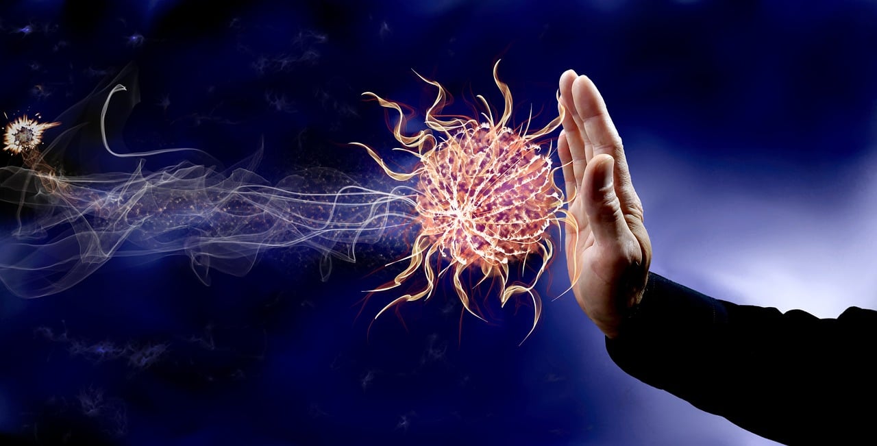
Protection of the organs most often affected by immune-related inflammation. The goal is to reduce overactive immune attacks on healthy organs without using broad-spectrum immunosuppressive medication.
The goal is to spare healthy organs from overactive immune attacks without blocking the treatment effect [119, 123].
See also Reduce inflammation
See also Immune response and inflammation
See also Inflammation and cancer
Omega-3 (high dose)
Fatty acids that help reduce inflammation in the skin and joints.
High-dose Omega-3 means a daily intake of 3 g or more of the active fatty acids EPA and DHA. This level is used to achieve a medical anti-inflammatory effect.
- Effect: Relieves immune-related side effects without being generally immunosuppressive.
- Interaction risk: Low. (primarily related to bleeding risk during surgery).
Article: [120] Fatty Acids as a Tool to Boost Cancer Immunotherapy Efficacy (ResearchGate, 2022)
- Content: A scientific review of the importance of fatty acids for the effect of immunotherapy; this is not a randomised study. The research documents that omega-3 (PUFA) helps preserve the patient’s body weight and muscle mass during the course of illness. The study shows that these fatty acids directly affect immune cells and have the potential to strengthen the clinical benefit of treatment through optimised nutritional status.
Boswellia Serrata
A potent herb for protecting the intestinal mucosa in immune-related inflammation.
- Effect: Reduces inflammation locally in the gut without suppressing the overall immune system.
- Half-life: ⬤ approx. 6 hours.
- Interaction risk: None known.
- Comment: Requires dosing 2–3 times daily for a stable effect.
Article: [123] Boswellia serrata Preserves Intestinal Epithelial Barrier from Oxidative and Inflammatory Damage (PLOS, 2015)
- Content: A scientific study of Boswellia’s ability to protect the intestinal barrier; this is not a randomised study. The research documents that the herb counteracts the breakdown of the proteins that seal the intestinal mucosa when exposed to inflammation. The study shows that Boswellia acts as an antioxidant that protects gut integrity and is therefore suitable as safe supportive care to prevent damage to the intestinal wall.
Lactobacillus rhamnosus GG (LGG)
Well-documented probiotic for preventing immune-related diarrhoea.
- Effect: Stabilises the intestinal barrier and reduces the need for immunosuppressive medication.
- Half-life: ◯ Continuously excreted.
- Interaction risk: None.
Article: [124] Gut microbiome on immune checkpoint inhibitor therapy and consequent immune-related colitis: a review (PubMed, 2023).
- Content: A scientific review of the link between the gut microbiome and side effects of immunotherapy; this is not a randomised study. The research documents that manipulating the gut flora via probiotics can be an effective method to reduce the risk of immune-related colitis. The study shows that balancing the gut flora is a practical way to protect the patient from toxic reactions that might otherwise necessitate stopping cancer treatment.
Melatonin
Helps target the immune system and protect healthy cells from oxidative damage.
- Effect: Regulates the immune response and reduces toxicity to the body’s healthy tissues. Weakens cancer cells’ ability to “hide” from the immune system by lowering PD-L1 levels (the cancer cells’ shield).
- Half-life: ◯ 30–50 minutes.
- Interaction risk: Low
- Comment: Should be taken in the evening to support the circadian rhythm.
Article: [125] Therapeutic Potential of Melatonin Counteracting Chemotherapy-Induced Toxicity in Breast Cancer Patients: A Systematic Review (MDPI, 2023)
- Content: A scientific review of melatonin’s ability to counteract side effects in breast cancer patients; this is not a randomised study. The research documents that doses of 20 mg daily increase the rate of partial response and 1-year survival. The study shows that the combination of melatonin and standard chemotherapy significantly improves patients’ quality of life and has a high safety profile as supportive care.
Article: [125A] Melatonin suppresses PD-L1 expression and exerts antitumor activity in hepatocellular carcinoma (Nature, 2025)
- Content: A scientific study of melatonin’s ability to inhibit cancer cells’ escape mechanisms; this is not a randomised study. The research documents that melatonin reduces PD-L1 expression in cancer cells, preventing them from deactivating the immune system. The study shows that melatonin both directly inhibits cancer cell growth while also increasing T-lymphocyte activity, making it a valuable support for immunotherapy.
Mitochondrial support (energy for T cells)
Treatment requires high metabolic energy for immune cells to fight the tumour effectively [126].
See also Metabolic principles in cancer research
See also About mitochondria – what are they
PQQ (Pyrroloquinoline quinone)
A micronutrient that optimises energy production specifically in white blood cells. It does not remove fatigue like a cup of coffee, but rather works as a slow recharge of the body’s batteries from the ground up.
- Effect: Counteracts T-cell “fatigue” (exhaustion) by stimulating the formation of new powerhouses and increasing their energy (ATP), so they retain the ability to attack cancer cells for longer.
- Half-life: ◯ Rapid excretion.
- Interaction risk: None.
Article: [126] Mitochondrial Metabolism in T-Cell Exhaustion (PubMed, 2025).
- Content: A scientific review of T-cell energy metabolism; this is not a randomised study. The research documents that chronic disease leads to immune exhaustion due to failing cellular energy production. The study shows that by stimulating the formation of new powerhouses (mitochondria) and increasing ATP production (the cell’s fuel), this exhaustion can be prevented and reversed, allowing cells to regain their ability to fight cancer.
Skin and mucous membranes

Protecting the body’s external and internal barriers is crucial to avoid treatment interruptions. When the immune system is overstimulated, it can attack healthy tissue, leading to painful sores and inflammatory conditions in the mouth and gastrointestinal tract [127]
See also Dry mouth, pain and mouth sores
See also Reflux and stomach ulcers/gastritis
Zinc-carnosine
A specific mineral compound that binds directly to mucosal damage and acts as a protective layer.
- Effect: Repair of the oral cavity and stomach in immune-related inflammation.
- Half-life: ◯ A few hours (primarily acts through local contact).
- Interaction risk: None.
Article: [127] A Review of Zinc-L-Carnosine and Its Positive Effects on Oral Mucositis, Taste Disorders, and Gastrointestinal Disorders (PubMed, 2020).
- Content: A scientific review of zinc-L-carnosine’s ability to repair damage in epithelial cells; this is not a randomised study. The research documents that, through local antioxidant and anti-inflammatory functions, the substance rebuilds mucous membranes in both the mouth and stomach. The study shows that the agent effectively prevents and treats mucositis (inflammation of the mucous membranes), which is crucial to avoid reduced quality of life and treatment interruptions.
Immune-related side effects
Protecting gut balance is crucial to avoid the inflammatory conditions that treatment can trigger.
Lactic acid bacteria and Bifido
The natural, beneficial bacteria that live in the gut and help regulate the immune system’s activity level.
- Effect: Restores balance in the gut flora and reduces local inflammatory reactions in the digestive system.
- Half-life: Not relevant (must be taken daily).
- Interaction risk: None.
Article: [129] The gut microbiota in cancer immunity and immunotherapy (Nature, 2025)
- Content: A scientific review of the interaction between the gut’s microorganisms and the immune system; this is not a randomised study. The research documents that beneficial bacteria such as lactic acid bacteria and bifido act as important allies that both enhance treatment and reduce the risk of side effects. The study shows that targeted use of these bacteria can restore balance in the gut and prevent cancer cells from escaping the immune system.
Conclusion
Support during immunotherapy is about creating the optimal conditions for the body’s own defences. By nurturing the microbiome and monitoring the body’s inflammatory response, you can both increase the likelihood that treatment works and reduce the risk of serious immune-related damage.
This requires precise timing, especially when immunotherapy is combined with traditional chemotherapy, to ensure that you neither block the immune system’s attack nor allow chemo toxicity to run out of control.
See also Safe measures during a cancer course
See also Integrative oncology
See also Quality of life and shared responsibility
See also Antioxidants – pros and cons
Links
[14] Herb-drug interactions in oncology: pharmacodynamic/pharmacokinetic mechanisms and risk prediction (PMC, 2025).
- Content: The scientific foundation for risk assessment and calculating washout periods.
[27] Clinical pharmacology—how it shapes the drug development journey (PMC, 2025)
- Content: The article explains how pharmacokinetic models and an understanding of half-lives are crucial for determining the correct timing and dose in oncology treatment pathways.
[29] Our defining research stories of 2025 (Cancer Research UK, 2025)
- Content: A summary of the year’s most important breakthroughs, including the importance of exercise for survival and new methods to predict chemo-resistance via DNA testing.
[118] A gut microbial signature for combination immune checkpoint blockade across cancer types (Nature, 2024)
- Content: Scientific study of the importance of bacterial strains; not randomised. Documents that specific microbes are crucial for the immune system’s ability to respond to PD-1 inhibitors and predict treatment success.
[119] Dual contribution of the gut microbiome to immunotherapy efficacy and toxicity: supportive care implications and recommendations (Research Gate, 2022)
- Content: Scientific study of the microbiome’s dual role; not randomised. The research documents a strong correlation between treatment efficacy and the risk of adverse events (IrAEs). The study confirms that the same bacteria that increase efficacy often also trigger inflammation, requiring a balanced strategy to avoid toxicity.
[120] Fatty Acids as a Tool to Boost Cancer Immunotherapy Efficacy (ResearchGate, 2022)
[121] Prebiotic Potential of Dietary Polyphenols in Colorectal Cancer Immunomodulation (MDPI, 2025)
[123] Boswellia serrata Preserves Intestinal Epithelial Barrier from Oxidative and Inflammatory Damage (PLOS, 2015)
[124] Gut microbiome on immune checkpoint inhibitor therapy and consequent immune-related colitis: a review (PubMed, 2023).
[125] Therapeutic Potential of Melatonin Counteracting Chemotherapy-Induced Toxicity in Breast Cancer Patients: A Systematic Review (MDPI, 2023)
[125A] Melatonin suppresses PD-L1 expression and exerts antitumor activity in hepatocellular carcinoma (Nature, 2025)
[126] Mitochondrial Metabolism in T-Cell Exhaustion (PubMed, 2025).
[127] A Review of Zinc-L-Carnosine and Its Positive Effects on Oral Mucositis, Taste Disorders, and Gastrointestinal Disorders (PubMed, 2020).
[128] Synergistic Effects of Polyphenols and Gut Microbiota (Wiley Online Library, 2026)
[129] The gut microbiota in cancer immunity and immunotherapy (Nature, 2025)
[130] Gut microbiota shapes cancer immunotherapy responses (Nature, 2025)
- Content: Scientific review of the role of gut bacteria; not randomised. The research documents that microbes such as Akkermansia recode the environment around cancer cells, enhancing treatment and reducing side effects.
[131] Steroid-sparing strategies for managing immune-related adverse events (Research Gate, 2026)
- Content: Scientific review of adverse-event management; not randomised. Documents that targeted support relieves inflammation without weakening the immune system’s ability to fight cancer, ensuring continuation of treatment.
Page created:
d. 11.02.26
❤
What you read on I Have Cancer is not a recommendation. Seek qualified guidance.
Biological half-lives – Links
Methodological basis for half-lives
To ensure clinical credibility, the specified values have been established through a hierarchical prioritization of data across four levels – under Biological half-lives – Links (at the bottom of the page) called Level of Evidence:
- 1. Human data
- The half-life is measured directly in human blood after ingestion of the substance or preparation.
- Precision: Very high. The figures are directly measurable and clinically verified.
- 2. Data for active ingredients
- The preparation itself has not been sufficiently studied in humans. The half-life is therefore based on one or more of the most important active substances in the preparation.
- Precision: Good. Measurements are based on the active drivers, even though the total product contains several components.
- 3. Calculated half-life
- No useful direct measurements are available. The half-life is estimated here based on knowledge of how the substance is absorbed, metabolized, and excreted.
- Precision: Based on biological plausibility and general biochemistry.
- 4. Preclinical data
- There is insufficient human data available. The half-life is based here on animal experiments or other preclinical data and is stated conservatively.
- Præcision: Vejledende. Her fungerer washout-perioden som en bevidst “over-sikring”.
By respecting these intervals, biosupport can be applied strategically during the recovery phase to optimize the overall course of treatment and minimize side effects without compromising the therapeutic index.
AHCC (active hexose correlated compound)
Washout: 2 days.
Level of Evidence: 2 (green).
- Half-life: ⬤ Estimated under 5 hours (plasma) / enzymatic impact normalized within 48 hours.
- Interaction risk Chemo: None known
- Interaction risk Radiation: None known
Documentation: AHCC is a fermented mushroom extract rich in acetylated alpha-glucans. A human Phase 1 study (Spierings et al., 2007) documents clinical safety at high doses. However, metabolic studies (Mach et al., 2008; Mathew et al., 2017) demonstrate that AHCC functions as both a substrate and inducer of the liver enzyme CYP2D6 (Phase 1) as well as an inducer of UGT 1A3 and 1A6 (Phase 2). As these enzyme pathways are responsible for the metabolism of many oncological drugs (e.g., tamoxifen and letrozole), a risk of reduced treatment efficacy exists. Based on this demonstrated enzymatic impact in human tissue, the washout period is set to 2 days to ensure normal metabolic conditions prior to treatment.
Link:
[A] Spierings E. L. et al.: A Phase I study of the safety of AHCC in healthy volunteers (J Nutr Sci Vitaminol, 2007) – Human safety.
[B] Mathew L., Gaikwad A., Smith J. A. et al.: Evaluation of Active Hexose Correlated Compound (AHCC) in Combination With Anticancer Hormones in Orthotopic Breast Cancer Models (Integrative Cancer Therapies, 2017)
[C] Coffer L. W. et al.: Evaluation of Active Hexose Correlated Compound (Ahcc) on Phase II Drug Metabolism Pathways and the Implications for Supplement-Drug Interactions (Semantic Scholar / ResearchGate, 2015)
Akkermansia (muciniphila)
Half-life: Not relevant as a classic plasma half-life; A. muciniphila is an intestinal bacterium and can colonize the gut after ingestion.
Evidence level: 4 (orange).
- Interaction risk Chemo: None known
- Interaction risk Radiation: None known
Documentation: Akkermansia muciniphila is an intestinal bacterium and therefore does not have a plasma half-life in the same way as a drug or an absorbed dietary supplement. Human studies show that oral administration can increase the presence of A. muciniphila in the gut, but there is insufficient human data to determine how long the bacterium or its biological impact persists after discontinuation. Therefore, a documented washout period cannot be calculated either.
No documentation has been found that A. muciniphila reduces the efficacy of chemotherapy. On the contrary, a preclinical study with cisplatin showed that the combination with A. muciniphila inhibited tumor growth more than cisplatin alone. However, this is animal data and cannot be used as documentation for the same effect in humans.
In radiotherapy, there are conflicting preclinical results regarding radiation damage to normal intestinal tissue, but no documentation of a reduced antitumor effect. A study found that A. muciniphila protected the intestine against radiation-induced damage, partly via propionic acid, while another study found increased acute intestinal damage under specific experimental conditions. The results thus relate to intestinal toxicity and cannot in themselves be characterized as an interaction with the cancer-killing effect of the radiotherapy.
Link:
[A] Depommier et al.: Supplementation with Akkermansia muciniphila in overweight and obese human volunteers: a proof-of-concept exploratory study (Nature Medicine, 2019)
- Content: Human intervention study documenting oral use and the impact on the presence of A. muciniphila in the gut. The study does not establish a half-life or washout after discontinuation.
[B] Chen et al.: Akkermansia muciniphila Enhances the Antitumor Effect of Cisplatin in Lewis Lung Cancer Mice (Journal of Immunology Research, 2020)
- Content: Animal study in which A. muciniphila in combination with cisplatin resulted in greater inhibition of tumor growth than cisplatin alone.
[C] He et al.: Akkermansia muciniphila protects the intestine from irradiation-induced injury by secretion of propionic acid (Gut Microbes, 2023)
- Content: Preclinical study with supplementary patient observations. In mice, A. muciniphila reduced radiation-induced intestinal damage; the effect was linked to propionic acid. The study does not investigate the protection of tumor cells against radiation.
[D] Wang et al.: Akkermansia muciniphila exacerbates acute radiation-induced intestinal injury by depleting mucin and enhancing inflammation (The ISME Journal, 2025)
- Content: Preclinical study in which A. muciniphila exacerbated acute radiation-induced intestinal damage under specific experimental conditions. The result relates to normal intestinal tissue and does not document a reduced antitumor effect of radiotherapy.
Activated charcoal
Washout: 1 day (deviation: based exclusively on gastrointestinal passage).
Level of Evidence: 1 (white).
- Half-life: Not relevant (not absorbed systemically, excreted via the intestine).
- Interaction risk Radiation: None known
Documentation: Since activated charcoal is not absorbed into the blood but remains in the gastrointestinal tract, its presence is governed exclusively by gastrointestinal transit time. According to the clinical status report (Silberman et al., 2023), the substance is most effective within 1 hour after ingestion, and its excretion follows the body’s natural passage (typically 12–24 hours). As there is no systemic half-life to account for, the washout period is based solely on ensuring complete passage through the gut before oncological treatment.
Link:
[A] Activated Charcoal (NIH, 2023)
Alpha-Lipoic Acid (ALA)
Washout: 2 days.
Level of Evidence: 1 (white).
- Half-life: ◯ approx. 15-60 minutes
- Interaction risk Chemo: Low
- Interaction risk Radiation: None known
Documentation: ALA is a sulfur-containing fatty acid that functions as a potent antioxidant in both aqueous and lipid phases. Human Phase 1 studies (Zárate et al., 2025) and randomized clinical trials (Yoon et al., 2016) unequivocally document that ALA is rapidly absorbed (tmax < 1 hour) and promptly eliminated from plasma with a half-life of less than 20 minutes for the biologically active R-form. Previous assumptions of long terminal elimination (based on preclinical models) have not been reproduced in human pharmacokinetic measurements over 36 hours. Since ALA effectively regenerates other antioxidants such as vitamins C and E, the washout period is conservatively set at 2 days to ensure that the enhanced antioxidant status is normalized before oncological treatment.
Link:
[A] Zárate E., Bravo-Lamicq C. et al.: Pharmacokinetics and safety of a fixed-dose combination of pregabalin and thioctic acid in healthy volunteers (Frontiers in Pharmacology, 2025) – Human safety. Latest human Phase 1 data.
[B] Yoon J., Moon S. J. et al.: Comparison of R(+)-α-lipoic acid exposure in healthy Korean male subjects (Translational and Clinical Pharmacology, 2016) – Documentation for the very short human half-life.
[C] Superti F. & Russo R.: Alpha-Lipoic Acid: Biological Mechanisms and Health Benefits (Antioxidants, 2024) – Systematic review of mechanisms of action.
Amygdalin (B17)
Washout: 2 days (deviation: requires time for elimination of toxic cyanide metabolite).
Level of Evidence: 1 (white).
- Half-life: ◯ 30-90 minutes (for the substance itself), but with a risk of cyanide accumulation.
- Interaction risk Chemo: Moderate
- Interaction risk Radiation: Moderate
Documentation: The clinical study (Moertel et al., 1982) conducted on 178 patients documents that amygdalin has no therapeutic effect on cancer but instead carries a significant risk of cyanide poisoning. The study showed that several patients had blood cyanide levels measured close to the lethal range. Since the substance is directly toxic and affects cellular oxygen uptake, the washout period is set at 2 days to ensure that cyanide levels have normalized before oncological treatment.
Link:
[A] A Clinical Trial of Amygdalin (Laetrile) in the Treatment of Human Cancer (The New England Journal of Medicine, 1982)
Andrographis paniculata
Washout: 2 days.
Evidence level: 1 (white)
- Half-life: ◯ approx. 2–3 hours for andrographolide; a few human studies find up to approx. 5 hours.
- Interaction risk Chemo: Moderate
- Interaction risk Radiation: Moderate
Documentation: Human pharmacokinetic studies following oral Andrographis paniculata show rapid absorption and a relatively short elimination half-life for andrographolide. Recent studies with standardized extracts typically find approx. 2.5–3 hours, while other human measurements range around 1–5 hours depending on dose and formulation. Andrographolide can simultaneously affect the response to chemotherapy. Preclinical studies show a synergistic effect together with cisplatin in several cancer models, including oral, lung, ovarian, and colon cancer, partly through increased apoptosis and impact on autophagy, ROS, and STAT3. For radiotherapy, there is direct preclinical documentation of radiosensitization; andrographolide increased radiation sensitivity both in vitro and in vivo through, among other things, inhibition of Akt and NF-κB signaling. Due to the short human half-life, but documented biological impact on both chemo and radiation responses, a conservative safety margin of 2 days is applied. See also Andrographis under Blood-Brain Barrier
Link:
- Content: Human pharmacokinetic study following oral Andrographis paniculata. The half-life of andrographolide was around 1–5 hours depending on dose and metabolite.
- Content: Human study of oral standardized extract. Andrographolide had a half-life of around 2.5–2.8 hours with single and repeated dosing.
- Content: Preclinical study in which andrographolide and cisplatin synergistically inhibited tumor growth and increased apoptosis.
[D] Synergistic antitumor effect of Andrographolide and cisplatin through ROS-mediated ER stress and STAT3 inhibition in colon cancer (Medical Oncology, 2022)
- Content: Preclinical study in which andrographolide enhanced the antitumor effect of cisplatin through increased ROS, ER stress, and STAT3 inhibition.
[E] Andrographolide sensitizes Ras-transformed cells to radiation in vitro and in vivo (2010)
- Content: Preclinical study documenting radiosensitization both in vitro and in vivo through inhibition of Akt and NF-κB signaling.
Apigenin
Washout: 19 days. (3 days for short-term use)
Level of Evidence: 4 orange (preclinical data and metabolic clearance).
- Interaction risk Chemo: Moderate
- Interaction risk Radiation: Moderate
Documentation: Apigenin is a flavonoid with complex pharmacokinetics. Systematic reviews of human data (Wang et al., 2019) report an average excretion half-life of approximately 12 hours, while the plasma half-life for free apigenin has been measured as low as 2.5 hours (DeRango-Adem et al., 2021). However, the classic kinetics study (Gradolatto et al., 2005) demonstrated a very slow elimination with a terminal half-life of 91.8 hours due to enterohepatic recirculation. Latest research (Sato et al., Nature 2024) confirms that modern nano-delivery systems significantly increase bioavailability, potentially increasing the risk of tissue accumulation. To ensure complete clearance during regular use, a safety interval of 19 days is maintained, while 3 days is considered sufficient for short-term use.
Link:
[A] Wang M., Firrman J. et al.: A Review on Flavonoid Apigenin: Dietary Intake, ADME, Antimicrobial Effects, and Interactions with Human Gut Microbiota (NIH, Biomed Res Int., 2019)
[B] Sato V. H., Sato H. et al.: Enhancement of in vitro transcellular absorption and in vivo oral bioavailability of apigenin by self-nanoemulsifying drug delivery systems (Scientific Reports, Nature, 2024)
[C] DeRango-Adem et al.: Does Oral Apigenin Have Real Potential for a Therapeutic Effect in the Context of Human Gastrointestinal and Other Cancers? (Frontiers in Pharmacology, 2021)
[D] Gradolatto et al.: PHARMACOKINETICS AND METABOLISM OF APIGENIN IN FEMALE AND MALE RATS AFTER A SINGLE ORAL ADMINISTRATION (Science Direct, 2005)
Artemisinin, Artemisea
Washout: 8 days (deviation due to sustained autoinduction of drug-metabolizing enzymes).
Evidence level: 1 (white) for artemisinin / 2 (green) for Artemisia annua extract.
- Half-life: ◯ approx. 90–260 minutes
- Interaction risk Chemo: Moderate
- Interaction risk Radiation: Moderate
Documentation: Human pharmacokinetic studies (Phase 1) following oral artemisinin show a short elimination half-life, typically approx. 90–160 minutes, but up to approx. 260 minutes depending on the study and dose (Duc et al., 1994; Benakis et al., 1997). The deciding factor for washout, however, is that artemisinin strongly induces its own metabolism and other CYP enzymes. Human studies show that this enzymatic impact can persist after the elimination of the substance itself; Gordi et al. (2005) estimated an enzyme half-life of approx. 38 hours and 6–8 days before enzyme activity normalizes. Furthermore, artemisinin can modify the effect of chemotherapy, including cisplatin and epirubicin, while preclinical studies show radiosensitization of cancer cells (Tran et al., 2014; Popova et al., 2022; Kim et al., 2012). The washout period is therefore set to 8 days for regular use. For Artemisia annua, the same safety margin is used, but evidence level 2, as the kinetics are extrapolated from the extract’s artemisinin content.
Link:
[A] Duc et al.: The pharmacokinetics of a single dose of artemisinin in healthy Vietnamese subjects (American Journal of Tropical Medicine and Hygiene, 1994)
- Content: Human pharmacokinetic study (Phase 1) following oral artemisinin with an average elimination half-life of approx. 155 minutes.
[B] Benakis et al.: Pharmacokinetics of artemisinin and artesunate after oral administration in healthy volunteers (American Journal of Tropical Medicine and Hygiene, 1997)
- Content: Human clinical study (Phase 1) of oral artemisinin with a terminal elimination half-life of approx. 260 minutes and rapid absorption and distribution.
[C] Gordi et al.: A semiphysiological pharmacokinetic model for artemisinin in healthy subjects incorporating autoinduction of metabolism and saturable first-pass hepatic extraction (British Journal of Clinical Pharmacology, 2005)
- Content: Human pharmacokinetic study (Phase 1) of repeated oral artemisinin. The enzymatic impact had an estimated half-life of approx. 38 hours, and the model showed that it takes around 6–8 days after the last dose before the induced enzyme activity returns to baseline.
[D] Mihara et al.: Stereospecific analysis of omeprazole supports artemisinin as a potent inducer of CYP2C19 (Fundamental & Clinical Pharmacology, 1999)
- Content: Human interaction study (Phase 1) documenting significant induction of drug metabolism after seven days of oral artemisinin. After a six-day washout, the measured enzyme-related values were normalized.
[E] Tran et al.: Comparative cytotoxicity of artemisinin and cisplatin and their interactions with chlorogenic acids in MCF7 breast cancer cells (Planta Medica, 2014)
- Content: Preclinical study of artemisinin and cisplatin, showing that the antitumor activity of artemisinin can be significantly altered by other plant components; particularly relevant when using the whole Artemisia annua extract.
[F] Popova et al.: Antitumor activity of the combination of artemisinin and epirubicin in human leukemia cells (Anticancer Agents in Medicinal Chemistry, 2022)
- Content: Preclinical study in which artemisinin and epirubicin showed synergistic antitumor effects, especially in doxorubicin-resistant leukemia cells.
[G] Kim et al.: Selective radiosensitization of human cervical cancer cells and normal cells by artemisinin through the abrogation of radiation-induced G2 block (International Journal of Radiation Oncology, Biology, Physics, 2012)
- Content: Preclinical study with artemisinin documenting increased radiation sensitivity in human cervical cancer cells by affecting the cells’ G2 checkpoint.
[H] Räth et al.: Pharmacokinetic study of artemisinin after oral intake of a traditional preparation of Artemisia annua L. (American Journal of Tropical Medicine and Hygiene, 2004)
- Content: Human pharmacokinetic study (Phase 1) of Artemisia annua tea, documenting systemic absorption of artemisinin from the whole plant and showing that absorption is faster than from tablets, while the overall bioavailability is comparable.
Ashwagandha
Washout: 3 days.
Evidence level: 1 (white).
- Half-life: ⬤ approx. 10 hours for withanolides in highly concentrated extract.
- Interaction risk Chemo: Moderate
- Interaction risk Radiation: Moderate
Documentation: Human pharmacokinetic studies (Phase 1) following oral Ashwagandha document systemic absorption of withanolides, but also show significant differences between extracts. In a randomized crossover study (Phase 1), withaferin A had a half-life of approx. 9.8 hours and withanolide A approx. 11 hours in a highly concentrated extract, while the values were significantly shorter in a weaker extract (Majeed et al., 2023). Withaferin A has marked biological activity in cancer cells, and preclinical studies describe both independent antitumor activity and interaction with conventional cancer treatment. In radiotherapy, the interaction is directly documented: withaferin A increased radiation sensitivity and cell death in experimental models (Devi et al., 1996), and recent research confirms radiosensitization through the impact on autophagy and mitochondrial function, among other things. As Ashwagandha extracts vary considerably in content and pharmacokinetics, and withanolides can affect the response to oncological treatment, a conservative safety margin of 3 days is applied.
Link:
[A] Majeed et al.: Pharmacokinetics and bioequivalence of Withania somnifera (Ashwagandha) extracts – A double blind, crossover study in healthy adults (Heliyon, 2023)
- Content: Randomized, double-blind human crossover study (Phase 1) following oral Ashwagandha. Documents large formulation-dependent differences in pharmacokinetics; in the highly concentrated extract, the half-life was approx. 9.8 hours for withaferin A and approx. 11 hours for withanolide A.
[B] Clinical pharmacokinetic evaluation of Withania somnifera (L.) Dunal root extract in healthy human volunteers (Journal of Ethnopharmacology, 2024)
- Content: Human study (Phase 1) in 18 healthy volunteers following a single oral dose of Ashwagandha root extract. Documents rapid absorption and measurable plasma concentrations of, among others, withanolide A and withaferin A.
[C] Devi et al.: Withaferin A: a new radiosensitizer from the Indian medicinal plant Withania somnifera (International Journal of Radiation Biology, 1996)
- Content: Preclinical study in which a non-toxic concentration of withaferin A increased radiation-induced cell death with a sensitizer enhancement ratio of 1.4–1.5.
[D] Lu et al.: Radiosensitizing effects of Withaferin A in gastric cancer cells via autophagy inhibition and mitochondrial disruption (Scientific Reports, 2025)
- Content: Preclinical study showing that withaferin A increases radiation sensitivity in gastric cancer cells through the impact on apoptosis, autophagy, and mitochondrial function.
Astragalus
Washout: 1 day.
Level of Evidence: 1 (white).
- Half-life: ◯ approx. 2.1–2.7 hours (human measurements).
- Interaction risk Chemo: Low
- Interaction risk Radiation: None known
Documentation: Astragalus contains saponins, with Astragaloside IV being the primary active marker. A comprehensive review (Stępnik et al., 2025) summarizes the substance’s anti-inflammatory and immunomodulatory effects. A human Phase 1 study (Xu et al., 2013) documents rapid and linear elimination in humans with a half-life of 2.1–2.7 hours and confirms that no accumulation occurs with daily dosing. The mathematical elimination (5 x t½) is thus completed in less than 14 hours. Preclinical models have shown half-lives of up to 5.5 hours (Tan et al., 2020), but these have not been reproduced in human trials. Since Astragalus has a low oral bioavailability of approximately 2.2% (ResearchGate, 2018) and the substance is excreted rapidly, a 24-hour washout ensures full elimination of the active components and a good margin for normalization of biological processes before oncological treatment.
Link:
[A] Xu M., Yin J. et al.: Pharmacokinetics and tolerance of total astragalosides after intravenous infusion in healthy Chinese volunteers (Phytomedicine, 2013) – Primary source for human kinetics.
[B] Stepnik et al.: In Vivo Insights into the Role of Astragaloside IV in Preventing and Treating Civilization Diseases: A Comprehensive Review (MDPI, 2025)
[C] Tan Y. Q. et al.: Astragaloside IV: An Effective Drug for the Treatment of Cardiovascular Diseases (Drug Des Devel Ther., 2020) – Preclinical data and comparison.
[D] Qing, et al.: Pharmacokinetics Comparison, Intestinal Absorption and Acute Toxicity of LS-102 (Research Gate, 2018) – Documentation for low oral absorption (2.2%) of AGS-IV.
Baicalin
Washout: 3 days.
Evidence level: 1 (white).
- Half-life: ⬤ approx. 4 to 11 hours.
- Interaction risk Chemo: Low
- Interaction risk Radiation: Low
Documentation: Human Phase 1 studies following oral baicalein show that the substance is rapidly converted to baicalin, which is the dominant metabolite in plasma. Baicalin had a terminal half-life of approx. 4.2–10.8 hours, while baicalein varied from approx. 1.9–15 hours depending on the dose (Li et al., 2014). With repeated dosing, moderate accumulation of both baicalein and baicalin is observed (Li et al., 2016). The pharmacokinetics are further complicated by the conversion between baicalein and baicalin, intestinal flora, and enterohepatic recirculation, and baicalin can affect co-administered drugs through metabolic enzymes and protein binding (Huang et al., 2019). Preclinical cancer studies show that baicalin/baicalein can alter the response to doxorubicin and cisplatin, among others, and research shows radiosensitization of cancer cells through the impact on JAK2/STAT3 signaling, among other things. Due to the human half-life, the recirculation, and documented impact on both chemo and radiation responses, a safety margin of 3 days is applied. See also Baicalin under Signal modulation and DNA protection
Link:
dose of baicalein chewable tablets in healthy subjects (Journal of Ethnopharmacology, 2014)
- Content: Randomized human Phase 1 study with 72 healthy participants. After oral baicalein, the terminal half-life of baicalin was measured at approx. 4.2–10.8 hours and baicalein at approx. 1.9–15 hours.
[B] Li et al.: Multiple-Ascending-Dose Pharmacokinetics and Safety Evaluation of Baicalein Chewable Tablets in Healthy Chinese Volunteers (Clinical Drug Investigation, 2016)
- Content: Human study (Phase 1) of repeated oral administration. Steady state was reached after repeated dosing, and the accumulation was approx. 1.7–2.1 times for baicalein and 1.7–2.5 times for baicalin.
[C] Huang et al.: Pharmacokinetics and Bioavailability Enhancement of Baicalin: A Review (European Journal of Drug Metabolism and Pharmacokinetics, 2019)
- Content: Pharmacokinetic review describing baicalin’s gastrointestinal conversion, enterohepatic recirculation, transport mechanisms, and complex metabolism, as well as the potential for drug interaction through metabolic enzymes and protein binding.
- Content: Review of oncological research. Summarizes preclinical documentation that baicalein and baicalin can increase the sensitivity of cancer cells to both chemotherapy and radiotherapy.
[E] Baicalein Enhances Radiosensitivity in Colorectal Cancer via JAK2/STAT3 Pathway Inhibition (2024)
- Content: Preclinical in vitro and in vivo study showing that baicalein increased radiation sensitivity in radioresistant colorectal cancer and enhanced the tumor-inhibitory effect of radiation through inhibition of JAK2/STAT3 signaling.
Berberine
Washout: 3 days.
Evidence level: 1 (white).
- Half-life: ⬤ approx. 3–6 hours for standard oral berberine; formulation can prolong elimination.
- Interaction risk Chemo: Moderate
- Interaction risk Radiation: Moderate
Half-life: ⬤ approx. 3–6 hours for standard oral berberine; formulation can prolong elimination. Interaction risk Chemo: Moderate Interaction risk Radiation: Moderate Documentation: Human pharmacokinetic studies (Phase 1) following oral berberine show a relatively short elimination half-life. A study with an oral Coptis formulation found approx. 5 hours, while earlier human data for standard berberine is around 3 hours. The formulation is of great importance: a human crossover study (Phase 1) showed that a liposomal/micellar formulation resulted in significantly higher exposure and slower elimination than standard berberine (Solnier et al., 2023). Berberine can simultaneously affect drug metabolism; in a human interaction study (Phase 1), berberine increased the exposure to cyclosporine, likely partly through inhibition of CYP3A4. In cancer treatment, preclinical data show that berberine can alter the response to several cytostatics, including cisplatin, doxorubicin, taxanes, and 5-FU, most often towards increased sensitivity, but the effect depends on the drug and model. In radiotherapy, there is also direct preclinical documentation of radiosensitization through, among other things, increased DNA damage and inhibited DNA repair, while other studies show radioprotection of normal tissue. Due to the human pharmacokinetics, formulation-dependent exposure, and documented impact on both drug metabolism and treatment response, a conservative safety margin of 3 days is applied.
Link:
Berberine Formulation with Enhanced Absorption In Vitro and in Human Volunteers (Pharmaceutics, 2023)
- Content: Human crossover study (Phase 1) following 500 mg oral berberine. Standard berberine had low systemic exposure, while a liposomal/micellar formulation resulted in approximately six times higher AUC and slower elimination. The article also refers to a human elimination half-life for standard berberine of approx. 3 hours.
- Content: Human pharmacokinetic study (Phase 1) following an oral berberine-containing Coptis formulation. The measured elimination half-life for berberine was around 5 hours.
[C] Xin et al.: The effects of berberine on the pharmacokinetics of cyclosporin A in healthy volunteers (Methods and Findings in Experimental and Clinical Pharmacology, 2006)
- Content: Human interaction study (Phase 1) in which berberine increased exposure to cyclosporine. The results support a clinically relevant impact on drug metabolism, partly via CYP3A4.
[D] Devarajan et al.: Berberine – A potent chemosensitizer and chemoprotector to conventional cancer therapies (Phytomedicine, 2021)
- Content: Review of berberine in combination with chemotherapy. Describes both chemosensitization and protection of normal tissue with several cytostatics and emphasizes that berberine can biologically alter the treatment response.
[E] Radiosensitization effects of berberine on human breast cancer cells (International Journal of Molecular Medicine, 2012)
- Content: Preclinical study in which berberine increased the sensitivity of breast cancer cells to ionizing radiation through cell cycle impact and inhibition of RAD51-mediated DNA repair.
[F] Radiotherapy Enhancing and Radioprotective Properties of Berberine: A Systematic Review (2024)
- Content: Systematic review showing berberine’s dual role: radiosensitization of cancer cells and radioprotection of normal tissue through various mechanisms.
Boron
Washout: 5 days.
Evidence level: 1 (white).
- Half-life: ⬤ approx. 21 hours.
- Interaction risk Chemo: None known
- Interaction risk Radiation: None known
Documentation: Human pharmacokinetic data (Phase 1) following oral administration show an elimination half-life for boron, primarily present as boric acid in plasma, of approx. 21 hours (Schou et al., 1984). Boron is efficiently absorbed, not metabolized to a significant degree, and excreted mainly unchanged through the kidneys; almost complete excretion is observed within 96 hours. No clinically relevant drug interactions have been demonstrated with standard oral boron supplementation, and the NIH states no known clinically relevant interactions with medications. At high pharmacological concentrations, however, boric acid can affect redox processes and ferroptosis, and has preclinically enhanced the cytotoxicity of cisplatin and doxorubicin, among others; this has not been documented with standard supplement doses in humans (Corti et al., 2023). Nor is there documentation that standard boron supplementation counteracts radiotherapy; boron is used in completely different concentrations and formulations in boron neutron capture therapy (BNCT), which cannot be transferred to dietary supplements. Based on the human half-life, a safety margin of 5 days is applied.
Link:
[A] Schou et al.: Human pharmacokinetics and safety of boric acid (Archives of Toxicology Supplement, 1984)
- Content: Human pharmacokinetic study (Phase 1) with oral boric acid in adult men. The average elimination half-life was approx. 21 hours, and almost the entire absorbed amount was excreted in the urine within 96 hours.
[B] Murray: A comparative review of the pharmacokinetics of boric acid in rodents and humans (Biological Trace Element Research, 1998)
- Content: Pharmacokinetic review confirming an approx. 21-hour human half-life following both oral and intravenous exposure, as well as renal excretion without significant metabolism.
[C] NIH Office of Dietary Supplements: Boron – Health Professional Fact Sheet
- Content: The NIH’s professional review of boron as a dietary supplement. States that boron is not known to have clinically relevant interactions with medications.
[D] Corti et al.: Enhancement of ferroptosis by boric acid and its potential use as chemosensitizer in anticancer chemotherapy (BioFactors, 2023)
- Content: Preclinical study in which high pharmacological concentrations of boric acid enhanced ferroptosis and increased the cytotoxic effect of cisplatin and doxorubicin, among others. Documents biological interaction, but not a clinical interaction with standard boron supplementation.
Boswellia, Frankincense
Washout: 4 days.
Level of Evidence: 1 (white).
- Half-life: ⬤ approx. 6.8 hours (AKBA) / measurable in plasma up to 48 hours.
- Interaction risk Chemo: Low
- Interaction risk Radiation: None known
Documentation: Boswellia serrata contains active boswellic acids (BA), including AKBA, which function as potent inhibitors of inflammatory mediators (Roy et al., 2019). A human Phase 1 study (Kulkarni et al., 2021) in healthy volunteers documents an initial elimination half-life for AKBA of 6.8 hours. However, recent clinical measurements of both raw extract and formulated particles (Schmiech et al., 2024) demonstrate residual plasma concentrations up to 48 hours after ingestion, due to the substance’s lipophilic nature and slow release from tissue stores. To comply with the pharmacological standard for complete elimination (5 \times t½ in the terminal phase), the washout period is set at 4 days (96 hours). This ensures full clearance of both plasma and tissue stores as well as normalization of inflammatory signaling pathways (5-LOX) before oncological treatment.
Links:
[A] Kulkarni:Pharmacokinetics of solid lipid Boswellia serrata particles in healthy subjects (PubMed, 2021) – Primary source for human kinetics (AKBA).
[B] Schmiech et al.: Single-dose comparative pharmacokinetic/pharmacodynamic study of a micellar formulation versus a native Boswellia serrata dry extract in healthy volunteers (Science Direct, 2024) – Documentation for terminal phase and 48-hour detection.
[C] Roy N. K. et al.: An Update on Pharmacological Potential of Boswellic Acids against Chronic Diseases (Int. J. Mol. Sci., 2019) – Review of molecular targets and bioavailability.
Butyric acid
Washout: 1 day.
Level of Evidence: 1 (white).
- Half-life: ◯ Very short (½-14 minutes).
- Interaction risk Chemo: None known
- Interaction risk Radiation: None known
Documentation: Pharmacokinetic measurements in humans (Daniel et al., 1989) show that butyric acid is eliminated extremely rapidly from the blood. The elimination curve is divided into two phases, with an initial half-life of only 0.5 minutes followed by a phase of 13.7 minutes. Although preclinical models (Jung et al., 2021) utilize tributyrin (TB) as a “prodrug” to create a more stable release in the gut, human data confirm that the systemic presence is very short-lived. Due to this lightning-fast metabolic turnover, the washout period is set at 1 day to ensure complete elimination before oncological treatment.
Link:
[A] Daniel et al.: Pharmacokinetic study of butyric acid administered in vivo as sodium and arginine butyrate salts (ScienceDirect, 1989)
[B] Jung et al.: An efficient system for intestinal on-site butyrate production using novel microbiome-derived esterases (Springer Nature, 2021)
Cannabis (THC/CBD)
Washout: 28 days (exception: based on slow release from adipose tissue and influence on CYP450).
Level of Evidence: 1 (white).
- Interaction risk Chemo: Low
- Interaction risk Radiation: None known
Documentation: The pharmacokinetics of cannabis are complex due to the substances’ high lipid solubility, which leads to extensive storage in the body’s adipose tissue. For THC, review studies (Huestis, 2007) document that the terminal half-life is between 20 and 30 hours, as the substance is slowly released from tissue stores into the blood. For CBD, recent pharmacokinetic modeling (Kolli et al., 2025) demonstrates that the terminal elimination half-life in humans is extremely long, exceeding 134 hours (>5.5 days), meaning it can take over 70 days to reach a steady state in the body. Since both substances affect the liver’s enzyme systems (especially CYP450) and have prolonged biological activity, the washout period is conservatively set at 4 weeks (28 days) to ensure complete elimination from tissue stores before oncological treatment.
Link:
[A] Lucas et al.: The pharmacokinetics and the pharmacodynamics of cannabinoids (British Journal of Clinical Pharmacology, 2018)
[B] Huestis: Human Cannabinoid Pharmacokinetics (Chemistry & Biodiversity, 2007)
[C] Kolli et al.: Cannabidiol Bioavailability Is Nonmonotonic with a Long Terminal Elimination Half-Life (Cannabis and Cannabinoid Research, Mary Ann Liebert, 2025)
CoQ10
Washout: 7 days.
Evidence level: 1 (white).
- Interaction risk Chemo: Moderate
- Interaction risk Radiation: Moderate
Documentation: Human pharmacokinetic studies (Phase 1) show that CoQ10 is absorbed slowly after oral ingestion and has a long elimination half-life of around 33 hours. The substance is highly lipophilic, is distributed to tissues, and is transported mainly in lipoproteins. At 5 × t½, the calculated elimination would be around 7 days. Due to the lipophilic nature and tissue distribution of CoQ10, a longer, conservative washout of 12 days is used.
CoQ10 functions as an electron carrier in the mitochondrial respiratory chain and as a lipid-soluble antioxidant. This provides a theoretical possibility of affecting cancer treatments where oxidative damage contributes to the therapeutic effect. However, clinical documentation that CoQ10 reduces the efficacy of chemotherapy is lacking. A systematic review of antioxidant supplementation during cancer treatment found insufficient evidence to determine whether CoQ10 affects treatment response or survival. Due to the antioxidant effect and the lack of sufficient clinical interaction studies, the risk is conservatively assessed as moderate.
The same issue applies to radiotherapy, where reactive oxygen species are a significant part of the biological effect of the radiation. There is no clinical documentation that CoQ10 protects tumor cells from radiation, but neither is there sufficient documentation to safely rule out such an impact. The interaction risk is therefore assessed as moderate.
Link:
[A] Tomono et al.: Pharmacokinetic study of deuterium-labelled coenzyme Q10 in man (International Journal of Clinical Pharmacology, Therapy, and Toxicology, 1986)
- Content: Human pharmacokinetic study (Phase 1) with isotope-labeled CoQ10 in 16 healthy men. Terminal elimination half-life 33.19 ± 5.32 hours and a secondary plasma peak after around 24 hours.
[B] Roffe et al.: Efficacy of coenzyme Q10 for improved tolerability of cancer treatments: a systematic review (Journal of Clinical Oncology, 2004)
- Content: Systematic review of six controlled clinical trials with CoQ10 in cancer patients, mainly during anthracycline treatment. There were signs of reduced treatment-related toxicity, but the evidence was insufficient for definitive conclusions.
[C] Yasueda et al.: Efficacy and Interaction of Antioxidant Supplements as Adjuvant Therapy in Cancer Treatment: A Systematic Review (Integrative Cancer Therapies, 2016)
- Content: Systematic review of clinical trials with antioxidants during cancer treatment, including CoQ10. Investigates both treatment toxicity and potential impact on antitumor effect.
[D] Nakayama et al.: Systematic Review: Generating Evidence-Based Guidelines on the Concurrent Use of Dietary Antioxidants and Chemotherapy or Radiotherapy (Cancer Investigation, 2011)
- Content: Systematic review of 52 clinical trials with antioxidants during chemotherapy and/or radiotherapy, including clinical studies with CoQ10. Highlights the limited documentation on whether antioxidant supplementation affects the treatment effect.
DCA (dichloroacetat)
Washout: 3 days.
Evidence level: 1 (white).
- Interaction risk Chemo: Moderate
- Interaction risk Radiation: Moderate
Documentation: Human pharmacokinetic studies (Phase 1) show a half-life for oral DCA of approx. 95 minutes after the first dose, but DCA inhibits its own metabolizing enzyme GSTZ1, so the half-life is significantly prolonged with repeated use and has been measured at an average of 3.6–9.9 hours (Curry et al., 1991). Human cancer studies confirm that kinetics are affected by repeated treatment and individual GSTZ1 variants (Tian et al., 2019). Furthermore, DCA can affect the response to conventional treatment. A randomized Phase 2 study has investigated DCA in combination with cisplatin and radiotherapy without demonstrated impairment of the treatment outcome, while preclinical studies show both synergistic and conflicting effects in radiotherapy. Due to the self-inhibiting metabolism and documented impact on treatment response, a safety margin of 3 days is applied..
Link:
[A] Curry S. H. et al.: Disposition and pharmacodynamics of dichloroacetate (DCA) and oxalate following oral DCA doses (Biopharmaceutics & Drug Disposition, 1991)
- Content: Human pharmacokinetic study (Phase 1) measuring the oral DCA half-life at approx. 95 minutes after the first dose and showing prolongation to an average of 3.6–9.9 hours after repeated dosing.
[B] Tian et al.: GSTZ1 genotypes correlate with dichloroacetate pharmacokinetics and chronic side effects in multiple myeloma patients in a pilot phase 2 clinical trial (Pharmacology Research & Perspectives, 2019).
- Content: Human Phase 2 pilot study in multiple myeloma patients showing an association between GSTZ1 genotype, DCA concentrations, pharmacokinetics, and side effects.
[C] Phase II study of dichloroacetate in combination with chemoradiotherapy for unresected, locally advanced head and neck squamous cell carcinoma (Investigational New Drugs, 2022)
- Content: Randomized Phase 2 study of oral DCA in combination with cisplatin and radiotherapy; the combination was feasible without a demonstrated negative effect on survival.
[D] Dichloroacetate induces tumor-specific radiosensitivity in vitro but attenuates radiation-induced tumor growth delay in vivo (Strahlentherapie und Onkologie, 2013)
- Content: Preclinical study finding radiosensitization in vitro, but simultaneously an opposite effect in vivo, where DCA reduced the growth-inhibitory effect of radiotherapy on xenograft tumors.
[E] Dichloroacetate Radiosensitizes Hypoxic Breast Cancer Cells (International Journal of Molecular Sciences, 2020)
- Content: Preclinical study showing increased ROS formation and radiosensitization of hypoxic breast cancer cells following DCA treatment.
DIM (diindolylmethane)
Washout: 3 days.
Level of Evidence: 1 (white).
- Half-life: ⬤ approx. 4-8 hours.
- Interaction risk Chemo: Low
- Interaction risk Radiation: None known
Documentation: DIM is the primary acid condensation product of indole-3-carbinol (I3C). Human pharmacokinetic studies show that following oral ingestion, I3C is rapidly converted into DIM, among others, which can be measured directly in plasma (Reed et al., 2006). Direct human pharmacokinetic data show an elimination half-life for DIM of around 4 hours, while monohydroxylated DIM metabolites and their conjugates have a longer half-life of around 9 hours (Maier et al., 2021). Since the active metabolites persist longer than the parent compound, the washout is not based solely on DIM’s plasma half-life. A safety margin of 3 days is applied to ensure the elimination of both DIM and the longer-persisting metabolites prior to oncological treatment.
Link:
[A] Srikanth et al.: Unveiling the Multifaceted Pharmacological Actions of Indole-3-Carbinol and Diindolylmethane: A Comprehensive Review (Plants, 2025)
[B] Reed et al.: Single-Dose and Multiple-Dose Administration of Indole-3-Carbinol to Women: Pharmacokinetics Based on 3,3′-Diindolylmethane (Cancer Epidemiol Biomarkers Prev, 2006)
[C] Maier et al.: 3,3′-Diindolylmethane Exhibits Significant Metabolism after Oral Dosing in Humans (Drug Metabolism and Disposition, 2021).
EGCG (Green Tea)
Washout: 2 days.
Evidence level: 1 (white)
- Half-life: ◯ approx. 2–5½ hours.
- Interaction risk Chemo: Moderate
- Interaction risk Radiation: Moderate
Documentation: Human pharmacokinetic studies (Phase 1) show that EGCG is absorbed and eliminated relatively quickly after oral ingestion, with a terminal half-life of approx. 2–5½ hours. At high or repeated doses, elimination may be somewhat prolonged. EGCG can simultaneously affect the response to chemotherapy. Preclinical studies show both synergistic and antagonistic interactions depending on the specific cytostatic drug; among other things, increased sensitivity to cisplatin has been shown. Therefore, EGCG cannot generally be considered either protective or inhibitory during chemotherapy.
In radiotherapy, there are both preclinical and human data. A study in breast cancer patients showed biological changes consistent with increased radiation sensitivity, while EGCG simultaneously has antioxidative and tissue-protective properties. There is no documentation of a general reduction in the antitumor effect of radiotherapy, but because EGCG can biologically affect the treatment response, the interaction risk is assessed as moderate. Due to the short human half-life, but the possibility of pharmacodynamic interactions, a conservative safety margin of 2 days is applied.
Link:
[A] Ullmann et al.: A single ascending dose study of epigallocatechin gallate in healthy volunteers (Journal of International Medical Research, 2003)
- Content: Human pharmacokinetic study (Phase 1) with oral EGCG. Documents rapid elimination and a terminal half-life of a few hours.
[B] Wang et al.: EGCG Enhances Cisplatin Sensitivity by Regulating Expression of the Copper and Cisplatin Influx Transporter CTR1 in Ovary Cancer (PLoS One, 2015)
- Content: Preclinical study in which EGCG increased the uptake of cisplatin and the cancer cells’ sensitivity to the treatment, as well as enhanced tumor inhibition in a xenograft model.
[C] The interactions of anticancer agents with tea catechins: current evidence from preclinical studies (2014)
- Content: Scientific review of the interaction of tea catechins with anticancer drugs. Documents that EGCG, depending on the drug and experimental model, can have both synergistic and antagonistic effects.
[D] Zhang et al.: Anti-cancer activities of tea epigallocatechin-3-gallate in breast cancer patients under radiotherapy (Current Molecular Medicine, 2012)
- Content: Human study in breast cancer patients undergoing radiotherapy. EGCG was associated with changes in relevant tumor and signaling markers; supplementary experimental data showed increased radiation sensitivity.
Genistein
Washout: 3 days.
Evidence level: 1 (white).
- Half-life: ⬤ approx. 8–13 hours.
- Interaction risk Chemo: Moderate
- Interaction risk Radiation: Moderate
Documentation: Human pharmacokinetic studies (Phase 1) following oral genistein show terminal half-lives of approx. 8–13 hours depending on dose and measurement method (Ullmann et al., 2005; Zhou et al., 2014). With repeated dosing, no significant accumulation was found. Genistein also has a documented pharmacodynamic interaction with oncological treatment. Preclinical studies show increased apoptosis and antitumor effect together with cisplatin, docetaxel, and doxorubicin (Li et al., 2005), while genistein can increase the sensitivity of cancer cells to radiation (Hillman et al., 2001). Due to the human half-life and the documented impact on both chemo and radiation responses, a safety margin of 3 days is applied.
Link:
[A] Ullmann et al.: Safety, tolerability, and pharmacokinetics of single ascending doses of synthetic genistein (Bonistein) in healthy volunteers (Advances in Therapy, 2005)
- Content: Randomized human Phase 1 study following oral genistein. The average terminal half-lives were approx. 7.5–10.2 hours at doses of 30–300 mg.
[B] Zhou et al.: Single- and multiple-dose pharmacokinetics of genistein capsules in healthy Chinese subjects: A phase I, randomized, open-label study (Clinical Therapeutics, 2014)
- Content: Human pharmacokinetic study (Phase 1) with single and repeated oral genistein. The half-life was approx. 9–13 hours, and no significant accumulation was found after repeated dosing.
[C] Li et al.: Inactivation of nuclear factor kappaB by soy isoflavone genistein contributes to increased apoptosis induced by chemotherapeutic agents in human cancer cells (Cancer Research, 2005)
- Content: Preclinical in vitro and animal studies in which genistein increased growth inhibition and apoptosis induced by cisplatin, docetaxel, and doxorubicin through, among other things, inhibition of NF-κB.
[D] Hillman et al.: Genistein potentiates the radiation effect on prostate carcinoma cells (Clinical Cancer Research, 2001)
- Content: Preclinical study in which genistein enhanced the effect of ionizing radiation on prostate cancer cells, thus documenting a biologically relevant interaction with radiotherapy.
Ginseng (Panax ginseng / Korean red ginseng)
Washout: 14 days.
Evidence level: 2 (green).
- Interaction risk Chemo: Low
- Interaction risk Radiation: Moderate
Documentation: Ginseng is a complex extract with several pharmacologically active ginsenosides, which have very different half-lives. Human studies (Phase 1) following oral red ginseng show that Rb1 is among the longest-persisting components with a half-life of approx. 38–69 hours with single and repeated dosing, while the metabolite Compound K is eliminated in approx. 8–10 hours (Choi et al., 2020). Another human study (Phase 1) found an approx. 58-hour half-life for Rb1 (Kim, 2013). Repeated use leads to significant accumulation of Rb1, Rb2, and Rc. At the same time, ginseng has been investigated clinically in several chemotherapy regimens, including FOLFOX, gemcitabine, and 5-FU, with no signs of reduced treatment efficacy (Kim et al., 2020; Yoon et al., 2021). In radiotherapy, the basis is more uncertain because ginseng and ginsenosides have documented radioprotective and antioxidative properties in preclinical models. Due to the long human half-life of Rb1, accumulation with repeated use, and possible radioprotection, a safety margin of 14 days is applied.
Link:
[A] Choi et al.: Tolerability and pharmacokinetics of ginsenosides Rb1, Rb2, Rc, Rd, and compound K after single or multiple administration of red ginseng extract in human beings (Journal of Ginseng Research, 2020)
- Content: Human pharmacokinetic study (Phase 1) following single and repeated oral red ginseng. Rb1, Rb2, and Rc had long terminal half-lives of around 36–69 hours and accumulated 4.5–6.7 times with repeated administration.
[B] Kim: Pharmacokinetics of ginsenoside Rb1 and its metabolite compound K after oral administration of Korean Red Ginseng extract (Journal of Ginseng Research, 2013)
- Content: Human study (Phase 1) of oral Korean red ginseng. The measured half-life was approx. 58 hours for Rb1 and approx. 8 hours for Compound K.
[C] Lee et al.: Studies on absorption, distribution and metabolism of ginseng in humans after oral administration (Journal of Ethnopharmacology, 2009)
- Content: Human study (Phase 1) documenting the intestinal flora’s conversion of ginsenosides and the subsequent systemic absorption of Compound K following oral ginseng.
[D] Kim et al.: Korean red ginseng for cancer-related fatigue in colorectal cancer patients with chemotherapy: A randomised phase III trial (European Journal of Cancer, 2020)
- Content: Randomized, double-blind Phase 3 study with 438 patients, where Korean red ginseng was given concurrently with mFOLFOX-6 chemotherapy. The study provides direct human documentation for the concurrent use of ginseng and chemotherapy.
[E] Yoon et al.: Immune-modulating Effect of Korean Red Ginseng by Balancing the Ratio of Peripheral T Lymphocytes in Bile Duct or Pancreatic Cancer Patients With Adjuvant Chemotherapy (In Vivo, 2021)
- Content: Randomized clinical study (Phase 2) where red ginseng was used during 5-FU/leucovorin or gemcitabine treatment. No increased incidence of neutropenia or liver impact was found.
[F] Lee & Son: Radioprotective potential of ginseng (Mutagenesis, 2005)
- Content: Scientific review of preclinical studies documenting radioprotective properties of ginseng, including protection against radiation-induced DNA damage and possible antioxidative and immunomodulatory mechanisms.
Shark Liver Oil
Half-life: Not precisely established (incorporated into cell membranes and transformed into plasmalogens).
Washout: 14 days (exception: based on lipid remodeling in immune cells).
Level of Evidence: 3 (yellow).
- Interaction risk Chemo: Low
- Interaction risk Radiation: None known
Documentation: Shark liver oil is a rich source of alkylglycerols (AKG). Unlike many other preparations that merely circulate in plasma, AKG functions as building blocks incorporated into cell membranes. A phase 1 clinical trial (Paul et al., 2021) shows that shark liver oil supplementation for 3 weeks leads to a significant enrichment of plasmalogens in both plasma and white blood cells. The alkylglycerols themselves undergo extensive metabolic “remodeling” in the body. Review articles (Pugliese et al., 1998 & Iannitti & Palmieri, 2010) describe AKG as multifunctional with stimulating effects on macrophages and the immune system that can persist after ingestion. Since the preparation alters the lipid composition of immune cells over a prolonged period, and precise data for terminal elimination are lacking, the washout period is conservatively set at 14 days.
Link:
[A] Pugliese et al.: Some biological actions of alkylglycerols from shark liver oil (J Altern Complement Med, 1998)
[B] Iannitti & Palmieri: An Update on the Therapeutic Role of Alkylglycerols (Marine Drugs, 2010)
[C] Paul et al.: Shark liver oil supplementation enriches endogenous plasmalogens and reduces markers of dyslipidemia and inflammation (Journal of Lipid Research, 2021).
Honokiol, Magnolia extract
Half-life: 2.5–5 hours (in the elimination phase).
Washout: 3 days.
Level of Evidence: 4 (orange)
- Half-life: ◯ approx. 2.5-5 hours.
- Interaction risk Chemo: Low
- Interaction risk Radiation: None known
Documentation: There is insufficient human pharmacokinetic data to establish a plasma half-life for honokiol after oral administration. The available pharmacokinetic data are predominantly preclinical. Sarrica et al. (2018) review, among other things, an oral rat study in which honokiol had an elimination half-life of nearly 5 hours. Honokiol is mainly metabolized through glucuronidation and sulfation, and preclinical studies also show an impact on CYP enzymes, which can alter the metabolism of other drugs. Since human pharmacokinetics are lacking and there is potential interference with drug metabolism, the evidence is classified as level 4, and a conservative safety margin of 3 days is applied.
Link:
[A] Arora et al.: Honokiol: a novel natural agent for cancer prevention and therapy (Current Molecular Medicine, 2012)
[B] Sarrica et al.: Safety and Toxicology of Magnolol and Honokiol (Planta Medica, 2018)
[C] Kim et al.: Modulation of Rat Hepatic CYP1A and 2C Activity by
Honokiol and Magnolol: Differential Effects on
Phenacetin and Diclofenac Pharmacokinetics In Vivo (Molecules, 2018)
I3C (Indol-3-carbinol)
Washout: 3 days (exception: follows the elimination of the active metabolite DIM).
Level of Evidence: 1 (white).
- Half-life: ⬤ I3C undetectable in plasma after oral ingestion; DIM approx. 4 hours and DIM metabolites approx. 9 hours.
- Interaction risk Chemo: Low
- Interaction risk Radiation: None known
Documentation: I3C is an unstable molecule that, in the acidic environment of the stomach, rapidly converts into condensation products, especially DIM. In a human Phase 1 study, I3C itself could not be detected in plasma after oral administration, whereas DIM could be measured, and the pharmacokinetics were therefore evaluated based on this metabolite (Reed et al., 2006). Reed et al. (2008) subsequently investigated the pharmacokinetics of oral DIM directly in healthy subjects. Recent human data show a half-life of around 4 hours for DIM and around 9 hours for monohydroxylated DIM metabolites and their conjugates (Maier et al., 2021). The pharmacokinetic significance of I3C after oral ingestion is thus primarily tied to the active condensation products and their metabolites. On this basis, a washout period of 3 days is applied.
Link:
[A] Srikanth et al.: Unveiling the Multifaceted Pharmacological Actions of Indole-3-Carbinol and Diindolylmethane: A Comprehensive Review (PubMed, Plants, 2025)
[B] Reed et al.: Single-Dose and Multiple-Dose Administration of Indole-3-Carbinol to Women: Pharmacokinetics Based on 3,3′-Diindolylmethane (Cancer Epidemiol Biomarkers Prev, 2006)
[C] Reed et al.: Single-dose pharmacokinetics and tolerability of absorption-enhanced 3,3′-diindolylmethane in healthy subjects (Research Gate, Cancer Epidemiol Biomarkers Prev, 2008)
[D] Maier et al.: 3,3′-Diindolylmethane Exhibits Significant Metabolism after Oral Dosing in Humans (Drug Metabolism and Disposition, 2021).
Ginger
Washout: 2 days.
Level of Evidence: 1 (white).
- Half-life: ◯ approx. 0.6 to 2.4 hours.
- Interaction risk Chemo: Low
- Interaction risk Radiation: None known
Documentation: Ginger contains several active components, including gingerols and 6-shogaol. A phase 1 clinical trial (Zick et al., 2008) documents that these substances are absorbed rapidly but are primarily found as conjugated metabolites in plasma with a half-life of less than 2 hours. Another clinical trial (Zhang et al., 2022) confirms that the half-life for both the free substances and their metabolites remains stable between 0.6 and 2.4 hours, even with long-term use. A review article (Biomedicine & Pharmacotherapy, 2023) defines 10-gingerol as a central phenolic compound with significant anti-inflammatory properties and describes its role in managing complex physiological responses. Since the substances are metabolized and excreted rapidly without accumulation, the washout period is set at 2 days.
Link:
[A] Zick et al.: Pharmacokinetics of 6-, 8-, 10-Gingerols and 6-Shogaol and Conjugate Metabolites in Healthy Human Subjects (Cancer Epidemiol Biomarkers Prev, 2009)
[B] Zhang et al.: Pharmacokinetics of Gingerols, Shogaols, and Their Metabolites in Asthma Patients (J Agric Food Chem, 2023)
[C] ScienceDirect: 10-Gingerol – an overview (Biomedicine & Pharmacotherapy, 2023)
Inositol hexaphosphate (IP6 / phytic acid)
Washout: 2 days.
Evidence level: 1 (white).
- Half-life: ⬤ approx. 4 hours.
- Interaction risk Chemo: Moderate
- Interaction risk Radiation: None known
Documentation: Human studies show that oral IP6 can be absorbed systemically, and Grases et al. (2001) document absorption and subsequent excretion after oral administration. IP6 is simultaneously degraded into lower inositol phosphates and myo-inositol. Pharmacodynamically, IP6 has been investigated together with multiple cytostatics in preclinical models, where, among other things, a synergistic effect with doxorubicin, tamoxifen, and oxaliplatin has been reported, as well as an impact on acquired oxaliplatin resistance. In a human study of breast cancer patients undergoing adjuvant chemotherapy, myo-inositol was administered orally, while IP6 was used topically. The study can therefore not be used as documentation for concurrent oral IP6 during chemotherapy. There are insufficient data regarding a specific interaction with therapeutic ionizing radiation. On this basis, a washout period of 2 days is applied.
Link:
[A] Grases et al.: Absorption and excretion of orally administered inositol hexaphosphate (IP6 or phytate) in humans (BioFactors, 2001)
- Content: Human pharmacokinetic study (Phase 1) in healthy volunteers documenting systemic absorption of oral IP6, maximum plasma concentration after approx. 4 hours, and subsequent renal excretion.
[B] NCATS: Phytic Acid – Pharmacokinetic Data (National Center for Advancing Translational Sciences)
- Content: Pharmacokinetic human data for oral phytic acid/IP6 with a stated plasma half-life of approx. 4 hours following a single dose of 1,400 mg.
[C] Shamsuddin et al.: Inositol hexaphosphate (IP6) enhances the anti-proliferative effects of adriamycin and tamoxifen in breast cancer (Anticancer Research, 2003)
- Content: Preclinical study showing a synergistic effect between IP6 and both doxorubicin (Adriamycin) and tamoxifen in breast cancer cells, including drug-resistant cell lines.
[D] Liao et al.: Inositol hexaphosphate sensitizes hepatocellular carcinoma to oxaliplatin relating inhibition of CCN2-LRP6-β-catenin-ABCG1 signaling pathway (Journal of Cancer, 2021)
- Content: Preclinical in vitro and in vivo study showing a synergistic antitumor effect between IP6 and oxaliplatin, and increased sensitivity to oxaliplatin.
[E] Xu et al.: Inositol hexaphosphate enhances chemotherapy by reversing senescence induced by persistently activated PERK and diphthamide modification of eEF2 (Cancer Letters, 2024)
- Content: Preclinical study showing that IP6 can reverse acquired chemoresistance to oxaliplatin through the impact on PERK signaling and cellular senescence.
- Content: Human study in breast cancer patients receiving adjuvant chemotherapy. Myo-inositol was administered orally, while IP6 was used topically. The study can therefore be used as documentation for the clinical use of inositol/IP6 in connection with chemotherapy, but not as documentation for concurrent oral administration of IP6.
L-Carnitine and Acetyl-L-Carnitine, ALC
Washout: 14 days.
Evidence level: 1 (white).
- Interaction risk Chemo: Moderate
- Interaction risk Radiation: None known
Documentation: A human pharmacokinetic study (Phase 1) following oral ingestion measured an elimination half-life of 60.3 ± 15.0 hours for L-carnitine and 35.9 ± 28.9 hours for acetyl-L-carnitine. Carnitine is further distributed between different tissue pools, where muscle tissue in particular has a slow turnover. Human kinetic data describe a total turnover time of around 38–119 hours. A washout of 14 days is therefore a conservative rounding based on the upper end of the documented human kinetics.
In chemotherapy, L-carnitine and ALC should not be assessed entirely the same. ALC has been investigated directly during taxane treatment in a large randomized study (Phase 3) with breast cancer patients. ALC did not prevent neuropathy and after 24 weeks resulted in significantly more chemotherapy-induced peripheral neuropathy; grade 3–4 neurotoxicity also occurred more frequently. This is a documented clinical impact on treatment toxicity, but the study did not show that ALC reduced the antitumor effect of the chemotherapy. Preclinical data further show that ALC can increase the antitumor effect of cisplatin in certain models. The interaction risk in chemotherapy is therefore assessed as moderate, primarily due to ALC during neurotoxic chemotherapy.
In radiotherapy, no documentation of a negative interaction has been found. A randomized clinical trial (Phase 2) in patients with head and neck cancer used L-carnitine concurrently with chemoradiotherapy and found less treatment-related toxicity, while the tumor response was comparable between the groups. This provides no basis for a known negative interaction with radiotherapy.
Link:
[A] Cao et al.: Comparison of pharmacokinetics of L-carnitine, acetyl-L-carnitine and propionyl-L-carnitine after single oral administration of L-carnitine in healthy volunteers (Clinical and Investigative Medicine, 2009)
- Content: Human pharmacokinetic study (Phase 1) with oral L-carnitine. The elimination half-life was approx. 60 hours for L-carnitine and approx. 36 hours for acetyl-L-carnitine.
[B] Rebouche: Kinetics, pharmacokinetics, and regulation of L-carnitine and acetyl-L-carnitine metabolism (Annals of the New York Academy of Sciences, 2004)
- Content: Review of human carnitine kinetics, including absorption, renal elimination, tissue distribution, and slow-turnover tissue pools.
[C] Hershman et al.: Randomized double-blind placebo-controlled trial of acetyl-L-carnitine for the prevention of taxane-induced neuropathy in women undergoing adjuvant breast cancer therapy (Journal of Clinical Oncology, 2013)
- Content: Randomized, double-blind, placebo-controlled study (Phase 3) with 409 evaluable breast cancer patients. ALC during taxane treatment increased peripheral neuropathy after 24 weeks and resulted in more cases of severe neurotoxicity.
[D] Pacilli et al.: Metabolic approach to the enhancement of antitumor effect of chemotherapy: a key role of acetyl-L-carnitine (Clinical Cancer Research, 2010)
- Content: Preclinical study in which ALC increased the sensitivity to cisplatin in p53-competent cancer cells and improved the antitumor effect of cisplatin in a mouse model.
[E] L-Carnitine to Reduce Chemoradiotherapy-Induced Toxicity in Head and Neck Cancer: A Randomized Study (2025)
- Content: Randomized clinical trial (Phase 2) with L-carnitine during concurrent chemoradiotherapy. Treatment-related toxicity was reduced, while the tumor response was comparable between the groups.
LDN (Low Dose Naltrexone)
Washout: 3 days.
Evidence level: 1 (white).
- Half-life: ⬤ approx. 4 hours for naltrexone / approx. 12 hours for 6β-naltrexol.
- Interaction risk Chemo: Low
- Interaction risk Radiation: None known
Documentation: Human pharmacokinetic studies (Phase 1) following oral naltrexone show a plasma half-life of approx. 4 hours for the parent compound and approx. 12 hours for the active main metabolite 6β-naltrexol. Five half-lives for the longest-persisting relevant metabolite correspond to approx. 60 hours, which is why the washout is rounded to 3 days.
In chemotherapy, the available documentation predominantly points to a neutral or possible synergistic effect. Preclinical studies show increased sensitivity to cisplatin, oxaliplatin, and docetaxel, among others, under specific dosage and time courses. Reviews therefore describe LDN as a possible adjuvant treatment to chemotherapy rather than as a known counteracting factor. However, the effect may depend on the dose and treatment sequence, and clinical data remain limited. The interaction risk is therefore assessed as low rather than none known.
In radiotherapy, only limited data exist. A randomized study (Phase 2) in patients with high-grade glioma used LDN concurrently with radiotherapy and temozolomide with no difference in the level of side effects compared to placebo, but the study was not designed to investigate the impact on the antitumor effect. Therefore, there is no documentation of a known negative interaction with radiotherapy.
Link:
[A] Meyer et al.: Bioequivalence, dose-proportionality, and pharmacokinetics of naltrexone after oral administration (Journal of Clinical Psychiatry, 1984)
- Content: Human pharmacokinetic study (Phase 1) following oral naltrexone. The half-life was approx. 4 hours for naltrexone and approx. 12 hours for the main metabolite 6β-naltrexol.
[B] Donahue et al.: Low-dose naltrexone suppresses ovarian cancer and exhibits enhanced inhibition in combination with cisplatin (Experimental Biology and Medicine, 2011)
- Content: Preclinical in vitro and in vivo study in which LDN inhibited ovarian cancer, and the combination with cisplatin resulted in greater tumor inhibition than the treatments alone.
[C] Low-Dose Naltrexone as an Adjuvant in Combined Anticancer Therapy (Cancers, 2024)
- Content: Review of LDN in combination with cancer treatment. Summarizes preclinical data, which predominantly point to a synergistic or adjuvant effect together with cytostatics, but emphasizes the need for clinical studies.
[D] Low-Dose Naltrexone: What is the Evidence? A Narrative Review (2026)
- Content: Review identifying a randomized study (Phase 2) with LDN concurrently with radiotherapy and temozolomide in high-grade glioma. No increased level of side effects was found compared to placebo.
L-Glutamine
Washout: 5 hours.
Evidence level: 1 (white).
- Half-life: ◯ approx. 60 minutes.
- Interaction risk Chemo: None known
- Interaction risk Radiation: None known
Documentation: Human pharmacokinetic data (Phase 1) following oral L-glutamine show rapid absorption and elimination with a terminal half-life of around 1 hour. The plasma concentration peaks rapidly after ingestion, and no significant accumulation has been demonstrated with repeated dosing. Based on 5 × t½, this corresponds to a washout of approx. 5 hours.
In chemotherapy, glutamine has been investigated in several clinical trials, especially for the prevention and relief of treatment-related mucositis. Systematic reviews and meta-analyses of randomized trials have shown no signs that glutamine reduces the antitumor effect of chemotherapy. The results regarding the prevention of mucositis vary between studies, but several analyses find a reduced incidence or severity.
In radiotherapy, oral glutamine has also been investigated clinically, especially in patients with head and neck cancer. Meta-analyses show a possible reduction of radiation-induced oral mucositis without documentation of reduced treatment efficacy. There is therefore no basis for a known negative interaction with either chemotherapy or radiotherapy.
Important note: Should be used with great caution and only in consultation with a doctor, as certain types of cancer can utilize glutamine as an energy source for growth.
Link:
[A] Sadaf et al.: A Population Pharmacokinetic Analysis of L-Glutamine Exposure in Patients with Sickle Cell Disease: Evaluation of Dose and Food Effects (Clinical Pharmacokinetics, 2024)
- Content: Human pharmacokinetic study (Phase 1) with repeated oral L-glutamine. Average terminal half-life approx. 1 hour, rapid absorption, and no demonstrated accumulation with repeated dosing.
- Content: Meta-analysis of 15 randomized trials with 988 cancer patients undergoing chemotherapy and/or radiotherapy. Glutamine reduced several measures of severe mucositis and treatment interruptions with no signs of reduced antitumor effect.
[C] Alsubaie et al.: Glutamine for prevention and alleviation of radiation-induced oral mucositis in patients with head and neck squamous cell cancer (Head & Neck, 2021)
- Content: Systematic review and meta-analysis of 11 randomized trials with 922 patients undergoing radiotherapy. Oral glutamine reduced the severity of mucositis and several treatment-related consequences.
- Content: Systematic review of oral glutamine during chemotherapy and/or radiotherapy. Most included studies found low toxicity and no signals of a negative impact on the oncological treatment.
Lion’s Mane (Hericium erinaceus), Lions Mane
Washout: 3 days.
Evidence level: 4 (orange).
- Half-life: ⬤ approx. 7–8 hours for erinacine A and erinacine S (oral animal data).
- Interaction risk Chemo: None known
- Interaction risk Radiation: None known
Documentation: There are no human pharmacokinetic studies establishing the plasma half-life of Lion’s Mane or its main active compounds. The best available oral pharmacokinetic data come from rats: erinacine A had a half-life of approx. 490 minutes, while erinacine S had a half-life of approx. 440 minutes following oral administration of Hericium erinaceus mycelium (Hu et al., 2019; Li et al., 2021). Erinacine A could still be detected in certain tissues after 24 hours. In vitro studies with human liver tissue also show a metabolic half-life for erinacine A of approx. 115 minutes, but this cannot be equated with human plasma half-life. Preclinical studies of Lion’s Mane and erinacines exist as potential antitumor compounds, but I find no documentation of a clinically relevant counteraction of chemotherapy or radiotherapy. Due to the lack of human pharmacokinetic data and the complex composition of the extract, a conservative safety margin of 3 days is applied.
Link:
[A] Hu et al.: Absolute Bioavailability, Tissue Distribution, and Excretion of Erinacine S in Hericium erinaceus Mycelia (Molecules, 2019)
- Content: Oral pharmacokinetic rat study with Hericium erinaceus mycelium. Erinacine S had a plasma half-life of approx. 440 minutes (7.3 hours) and an absolute oral bioavailability of approx. 15%.
[B] Li et al.: Preclinical Bioavailability, Tissue Distribution, and Protein Binding Studies of Erinacine A, a Bioactive Compound from Hericium erinaceus Mycelia Using Validated LC-MS/MS Method (Molecules, 2021)
- Content: Oral pharmacokinetic rat study. Erinacine A had a plasma half-life of approx. 490 minutes (8.2 hours) and an oral bioavailability of approx. 24%. The substance could still be detected in the liver and heart, among other organs, after 24 hours.
[C] Identification of Common Liver Metabolites of the Natural Bioactive Compound Erinacine A, Purified from Hericium erinaceus Mycelium (Applied Sciences, 2022)
- Content: Metabolism study with rat and human liver material. The measured metabolic half-life for erinacine A was approx. 115 minutes in human liver S9, which documents human metabolic turnover, but not plasma half-life following oral ingestion.
[D] Key oncologic pathways inhibited by Erinacine A: A perspective for its development as an anticancer molecule (Biomedicine & Pharmacotherapy, 2023)
- Content: Scientific review of preclinical cancer studies with erinacine A. Describes the impact on several signaling pathways involved in proliferation, apoptosis, invasion, and metastasis, but provides no clinical documentation of interaction with chemotherapy or radiotherapy.
Liposomal Curcumin
Washout: 15 hours.
Level of Evidence: 1 (white).
- Half-life: ◯ 6–42 minutes (plasma) / up to 2 hours (tissue).
- Interaction risk Chemo: Low
- Interaction risk Radiation: Low
Documentation: Liposomal curcumin was developed to increase the systemic availability of curcumin because conventional oral curcumin has low water solubility, limited absorption, and extensive first-pass metabolism (Prasad et al., 2014). Direct human pharmacokinetics are available for intravenous liposomal curcumin. In a randomized, placebo-controlled phase I trial involving 50 healthy subjects, liposomal curcumin was administered intravenously at doses of 10–400 mg/m². Curcumin and the active metabolite tetrahydrocurcumin (THC) rapidly reached measurable plasma concentrations during the infusion but declined rapidly after the end of the infusion. The reported elimination half-life of curcumin was approximately 6–42 minutes depending on the dose, while the mean residence time was approximately 0.62 hours for curcumin and 1.69 hours for THC (Storka et al., 2015).
Curcumin is simultaneously distributed to cells and tissues. Bolger et al. (2017) demonstrated the uptake of liposomal curcumin and THC in human erythrocytes, mononuclear blood cells, and hepatocytes. The study thus supports cellular distribution and metabolism, but does not provide a basis for specifying a human tissue half-life. Curcumin is rapidly metabolized through reduction to, among other things, tetrahydrocurcumin and through phase II conjugation to glucuronide and sulfate metabolites.
The rapid elimination has also been investigated in cancer patients. In a phase I dose-escalation trial involving 32 patients with locally advanced or metastatic cancer, intravenous Lipocurc provided measurable plasma concentrations during infusion, followed by a rapid decline after the end of infusion (Greil et al., 2018). There is no clinical documentation showing that liposomal curcumin reduces the efficacy of chemotherapy or radiation therapy. Furthermore, liposomal curcumin has been investigated clinically in combination with conventional oncological treatment, and preclinical data predominantly describe chemo- and radiosensitizing rather than protective effects on tumor cells. Based on the current documentation, the interaction risk is therefore assessed as low for both chemotherapy and radiation therapy.
Direct human pharmacokinetics demonstrate very rapid elimination of intravenous liposomal curcumin. Even when based on the longest reported plasma half-life of approximately 42 minutes, 5 \times t½ corresponds to approximately 3.5 hours. Curcumin is taken up into blood cells and converted into, among other things, tetrahydrocurcumin as well as glucuronide and sulfate conjugates, but there is no documentation of a prolonged persistence necessitating a substantially longer safety margin. The washout period is set at 15 hours based on documented human plasma elimination.
Link:
[A] Storka et al.: Safety, tolerability and pharmacokinetics of liposomal curcumin in healthy humans (International Journal of Clinical Pharmacology and Therapeutics, 2015)
- Content: Randomized, placebo-controlled phase I trial involving 50 healthy subjects using intravenous liposomal curcumin. Documents direct human pharmacokinetics, including the rapid elimination of curcumin and THC after the end of infusion.
[B] Bolger et al.: Distribution and Metabolism of Lipocurc™ (Liposomal Curcumin) in Dog and Human Blood Cells: Species Selectivity and Pharmacokinetic Relevance (Anticancer Research, 2017).
- Content: Investigates the distribution and metabolism of Lipocurc and THC in human erythrocytes, mononuclear blood cells, and hepatocytes, among others, and documents cellular uptake of liposomal curcumin.
[C] Greil et al.: A phase 1 dose-escalation study on the safety, tolerability and activity of liposomal curcumin (Lipocurc™) in patients with locally advanced or metastatic cancer (Cancer Chemotherapy and Pharmacology, 2018).
- Content: Human phase I trial involving 32 cancer patients. Liposomal curcumin provided stable plasma levels during infusion, followed by a rapid drop to very low or undetectable concentrations after the end of infusion.
[D] Prasad et al.: Recent Developments in Delivery, Bioavailability, Absorption and Metabolism of Curcumin: the Golden Pigment from Golden Spice (Cancer Research and Treatment, 2014).
- Content: Comprehensive review of curcumin’s absorption, low bioavailability, metabolism, and various formulations for improving systemic exposure. Describes, among other things, reduction to tetrahydrocurcumin as well as glucuronide and sulfate conjugation.
[E] Holdhoff et al.: Phase I/II study of liposomal curcumin in combination with standard radiation and temozolomide in patients with newly diagnosed high-grade gliomas (Journal of Clinical Oncology, 2026, ASCO abstract).
- Content: Ongoing clinical phase I/II trial of intravenous liposomal curcumin concurrently with temozolomide and radiation therapy. Preliminary 2026 results describe the combination as feasible and generally well tolerated; the study is not yet sufficient to document a clinically synergistic effect.
Luteolin
Half-life: 2 days.
Washout: 2 days.
Level of Evidence: 4 (orange).
- Half-life: ⬤ approx. 5–9 hours after intravenous administration in rats; oral human t½ not established.
- Interaction risk Chemo: Low
- Interaction risk Radiation: Low
Documentation: There is insufficient human pharmacokinetic data to establish a secure elimination half-life for oral luteolin. Preclinical data show extensive metabolism and low oral bioavailability. In rats, Sarawek et al. (2008) measured a half-life of around 5–9 hours after intravenous administration. Following oral administration, plasma concentrations dropped so rapidly that the oral elimination half-life could not be reliably calculated. Luteolin is thus metabolized relatively quickly, but human oral elimination is not sufficiently characterized for a direct 5 x t½ calculation. On this basis, a washout period of 2 days is applied.
Link:
[A] Wang et al.: Progress, pharmacokinetics and future perspectives of luteolin modulating signaling pathways to exert anticancer effects: A review (Medicine (Baltimore), 2024)
[B] Lv et al.: Luteolin: exploring its therapeutic potential and molecular mechanisms in pulmonary diseases (Frontiers in Pharmacology, 2025)
[C] Sarawek et al.: Pharmacokinetics of Luteolin and Metabolites in Rats (Natural Product Communications, 2008)
Lysine
Washout: 1 days.
Level of Evidence: 1 (white).
- Half-life: ◯ 2,8 hours.
- Interaction risk Chemo: None known
- Interaction risk Radiation: None known
Documentation: Human pharmacokinetic data are available for oral L-lysine. Irving et al. (1986) followed isotopically labeled lysine after oral administration in healthy young women and described the plasma kinetics using a multi-compartment model. The slowest elimination constant after oral administration averaged 0.00412 min⁻¹, corresponding to a terminal half-life of approx. 2.8 hours. Lysine is simultaneously involved in the body’s normal amino acid and protein metabolism, including tissue uptake, protein synthesis, and oxidation. Based on the human terminal plasma half-life, 5 x t½ equals approx. 14 hours. The washout period is therefore conservatively set to 1 day.
Link:
[A] Holeček: Lysine: Sources, Metabolism, Physiological Importance, and Use as a Supplement (International Journal of Molecular Sciences, 2025)
[B] Irving et al.: Lysine and Protein Metabolism in Young Women: Subdivision Based on the Novel Use of Multiple Stable Isotopic Labels (Journal of Clinical Investigation, 1986).
[C] Bier: Amino Acid Pharmacokinetics and Safety Assessment (The Journal of Nutrition, 2003)
Magnesium
Washout: 2 days.
Evidence level: 1 (white).
- Half-life: ⬤ approx. 8 hours for oral magnesium hydroxide.
- Interaction risk Chemo: None known
- Interaction risk Radiation: None known
Documentation: Magnesium is an essential mineral with tight physiological regulation, and the pharmacokinetics are more difficult to describe than for a standard drug because magnesium is already present in blood, bones, and tissues. A human crossover study (Phase 1) with oral magnesium hydroxide measured an elimination half-life of approx. 8.3 hours. Other human studies (Phase 1) of oral magnesium salts show changes in serum and urine magnesium during the first 24 hours, but also emphasize that different forms of magnesium are absorbed differently.
No documentation has been found showing that magnesium reduces the efficacy of chemotherapy. On the contrary, magnesium supplementation is clinically relevant during cisplatin treatment, among others, because cisplatin can cause significant renal magnesium loss and hypomagnesemia. Magnesium has also been administered concurrently with cisplatin in clinical trials (Phase 2) without signs of a reduced antitumor effect.
Furthermore, no documentation of a negative interaction with radiotherapy has been found. The long biological turnover of magnesium in bone and tissue stores is not the same as sustained elevated free plasma concentrations and should therefore not be used to calculate washout. With a human plasma half-life of around 8 hours, 5 × t½ would be under 2 days; therefore, the general minimum washout of 2 days is applied.
Link:
[A] Siener et al.: Pharmacokinetic Profile of Oral Magnesium Hydroxide (Basic & Clinical Pharmacology & Toxicology, 2017)
- Content: Human crossover study (Phase 1) in healthy participants following oral magnesium hydroxide. The calculated elimination half-life was approx. 8.3 hours.
[B] White et al.: Blood and urinary magnesium kinetics after oral magnesium supplements (Clinical Therapeutics, 1992)
- Content: Randomized human crossover study (Phase 1) of several oral magnesium forms. Documents absorption and changes in serum, leukocyte, and urine magnesium over 24 hours.
[C] Ballmer & Reinhart: Does parenteral magnesium sulfate have an antiemetic effect during chemotherapy with cis-platinum? (Cancer Chemotherapy and Pharmacology, 1989)
- Content: Randomized clinical trial (Phase 2) in which magnesium was administered concurrently with cisplatin-based chemotherapy. The study provides direct human documentation for concurrent use.
[D] Memorial Sloan Kettering Cancer Center: Magnesium
- Content: Clinical professional overview of magnesium and drug interactions. Describes, among other things, hypomagnesemia with cisplatin and with EGFR inhibitors such as cetuximab and panitumumab.
Maitake (Grifola frondosa)
Washout: 3 days.
Note: Beta-glucans from medicinal mushrooms are large molecules that are generally not absorbed into the bloodstream, but instead activate immune cells locally in the intestinal wall. Although the substance passes relatively quickly through the digestive system, the biological activation of the immune cells persists. The washout period of 5 days is a conservative safety margin that ensures complete rest in the immune system before oncological treatment.
Evidence level: 4 (orange).
- Half-life: ◯ approx. 3 hours (preclinical data).
- Interaction risk Chemo: Low
- Interaction risk Radiation: Low
Documentation: There is no established human plasma half-life for Maitake extract or Maitake D-fraction. Preclinical pharmacokinetic data for orally administered Maitake beta-glucans state a terminal elimination half-life of approx. 3 hours. Maitake also has documented positive biological activity during oncological treatment. Animal studies show an increased antitumor effect together with both mitomycin-C and cisplatin, and preclinical studies find a similarly enhanced effect together with taxol, cisplatin, and FOLFOX. Furthermore, a randomized clinical trial (Phase 2) has used oral Maitake D-fraction during concurrent chemoradiotherapy without signs of counteracting the treatment. Due to the lack of human pharmacokinetics and documented biological interaction with conventional treatment, a conservative safety margin of 3 days is applied.
Link:
[A] Molecular Mechanism Induced by Beta-Glucans from Maitake to Recover T Cell-Subpopulations during Immunosuppression (IntechOpen, 2020)
- Content: Reviews pharmacokinetic studies of oral Maitake beta-glucan; the terminal elimination half-life was calculated to be approx. 2.9 hours.
[B] Kodama et al.: Maitake D-Fraction enhances antitumor effects and reduces immunosuppression by mitomycin-C in tumor-bearing mice (Nutrition, 2005)
- Content: Animal study in which Maitake D-fraction enhanced the antitumor effect of mitomycin-C and counteracted the chemotherapy-induced immunosuppression.
[C] Maitake β-glucan enhances therapeutic effect and reduces myelosupression and nephrotoxicity of cisplatin in mice (International Immunopharmacology, 2009)
- Content: Animal study in which Maitake beta-glucan enhanced the antitumor and antimetastatic effect of cisplatin and simultaneously reduced myelosuppression and nephrotoxicity.
[D] Hu & Xie: Effect of Maitake D-fraction in advanced laryngeal and pharyngeal cancers during concurrent chemoradiotherapy: A randomized clinical trial (Acta Biochimica Polonica, 2022)
- Content: Randomized clinical trial (Phase 2) in which oral Maitake D-fraction was given during concurrent chemoradiotherapy in patients with advanced head and neck cancer. The study found reduced treatment-related side effects and better quality of life.
[E] DeMarco et al.: Black Maitake mushroom (Grifola frondosa) extract exerts anti-cancer effects and enhances the efficacy of chemotherapeutic drugs in 2D and 3D models of triple-negative breast cancer (Journal of Complementary and Integrative Medicine, 2026)
- Content: Preclinical study in which Maitake extract enhanced the effect of both cisplatin and taxol in models of triple-negative breast cancer.
[F] Drew et al.: Evaluating the anticancer activity of black maitake odaira extract-prothera and its interactions with common chemotherapeutics in human colorectal cancers (Journal of Complementary and Integrative Medicine, 2026)
- Content: Preclinical study finding positive interactions between Maitake extract and FOLFOX, as well as increased antitumor activity in a xenograft model.
Milk Thistle (Silymarin/ Silybin)
Washout: 2 days.
Evidence level: 1 (white).
- Half-life: ◯ approx. 1–3 hours for free silymarin flavonolignans / approx. 3–8 hours for conjugated metabolites.
- Interaction risk – Chemotherapy: Low
- Interaction risk – Radiotherapy: None known
Documentation: Human pharmacokinetic studies show that the principal flavonolignans in silymarin are absorbed and eliminated relatively rapidly after oral administration. The half-life varies between the individual compounds and their conjugated metabolites but is predominantly within a few hours. Silybin undergoes extensive glucuronidation and sulfation and is excreted, among other routes, via the bile. Based on the human pharmacokinetic data, a conservative washout period of 2 days is used.
No general adverse clinical interaction with chemotherapy has been documented. In a human study in cancer patients, concomitant use of milk thistle had no significant effect on irinotecan clearance, although laboratory studies indicate that silybin may affect enzymes such as CYP3A4 and UGT1A1. The interaction risk is therefore considered low.
For radiotherapy, no evidence has been found that silymarin or silibinin protects cancer cells against the effects of treatment. On the contrary, systematic reviews show that silymarin/silibinin may increase radiosensitivity and enhance radiation-induced cell death in preclinical cancer models, while other studies demonstrate protection of normal tissue against radiation-induced damage. This interesting dual pattern has not yet been sufficiently documented clinically to recommend concomitant use, but it does not provide grounds for classifying the radiotherapy interaction risk as moderate.
Link:
-[A] Silybin, a Major Bioactive Component of Milk Thistle – Chemistry, Bioavailability, and Metabolism (Molecules, 2018)
- Content: Review of human pharmacokinetic data for silybin/silymarin, including absorption, metabolism, and half-lives of a few hours.
[B] van Erp et al.: Effect of Milk Thistle (Silybum marianum) on the Pharmacokinetics of Irinotecan (Clinical Cancer Research, 2005)
- Content: Human study in cancer patients. Milk thistle had no significant effect on irinotecan clearance despite preclinical concerns regarding CYP3A4 and UGT1A1 interactions.
[C] Latacela et al.: The Radioprotective Potentials of Silymarin/Silibinin Against Radiotherapy-Induced Toxicities (Current Medicinal Chemistry, 2023)
- Content: Systematic review of 19 studies. In most studies, silymarin/silibinin reduced radiation-induced damage in healthy cells and tissues.
[D] Gupta et al.: The Radiosensitizing Potentials of Silymarin/Silibinin in Cancer: A Systematic Review (Current Medicinal Chemistry, 2024)
- Content: Systematic review of cancer models. In most studies, silymarin/silibinin increased the sensitivity of cancer cells to radiation and was associated with increased DNA damage, apoptosis, and tumour inhibition. Clinical evidence remains lacking.
Melatonin
Washout: 1 day.
Evidence level: 1 (white).
- Half-life: ◯ approx. 45 minutes (typical interval approx. 28–126 minutes).
- Interaction risk Chemo: Low
- Interaction risk Radiation: Low
Documentation: Human pharmacokinetic studies show that melatonin is rapidly absorbed and eliminated. A systematic review of 22 studies found half-lives ranging from 28 to 126 minutes, typically around 45 minutes. A Danish crossover study found approximately 54 minutes after oral and 39 minutes after intravenous administration. The short half-life makes 1 day a conservative washout period with a substantial safety margin.
Melatonin has been investigated alongside chemotherapy in several randomized clinical trials and meta-analyses without evidence of reduced treatment efficacy. Some studies have instead reported improved tumour response and fewer treatment-related adverse effects. However, the evidence is methodologically heterogeneous, and a recent Cochrane review considers several clinical effects to remain uncertain. The interaction risk with chemotherapy is therefore conservatively assessed as low.
For radiotherapy, the clinical evidence is less extensive, but randomized trials and meta-analyses have not shown evidence of reduced tumour response. Preclinical studies also demonstrate radioprotective effects in normal tissue. Because the clinical evidence is more limited, the interaction risk is conservatively maintained as low.
Link:
[A] Andersen et al.: Pharmacokinetics of oral and intravenous melatonin in healthy volunteers (BMC Pharmacology and Toxicology, 2016)
- Content: Human crossover study. The elimination half-life was approx. 54 minutes after oral melatonin and approx. 39 minutes after intravenous administration; oral bioavailability was approx. 3%.
[B] Harpsøe et al.: Clinical pharmacokinetics of melatonin: a systematic review (European Journal of Clinical Pharmacology, 2015)
- Content: Systematic review of 22 human pharmacokinetic studies. The half-life varied from 28 to 126 minutes and was typically around 45 minutes.
[C] Seely et al.: Melatonin as adjuvant cancer care with and without chemotherapy: a systematic review and meta-analysis of randomized trials (Integrative Cancer Therapies, 2012)
- Content: Systematic review and meta-analysis of 21 randomized trials. Concurrent melatonin was not associated with reduced treatment efficacy and was in the included studies associated with better tumor response and fewer chemotherapy-related side effects.
[D] Wang et al.: The efficacy and safety of melatonin in concurrent chemotherapy or radiotherapy for solid tumors: a meta-analysis of randomized controlled trials (Cancer Chemotherapy and Pharmacology, 2012)
- Content: Meta-analysis of eight randomized trials with 761 patients, where melatonin was used concurrently with chemotherapy or radiotherapy. No signs of reduced tumor response were seen; the overall results showed higher tumor remission and fewer treatment-related side effects.
[E] Lissoni et al.: Decreased toxicity and increased efficacy of cancer chemotherapy using the pineal hormone melatonin in metastatic solid tumour patients with poor clinical status (European Journal of Cancer, 1999)
- Content: Randomized trial with 250 patients with metastatic solid tumors. Melatonin was given concurrently with several different chemotherapy regimens and was not associated with reduced treatment efficacy.
[F] Posadzki et al.: Melatonin in cancer treatment (Cochrane Database of Systematic Reviews, 2025)
- Content: Cochrane review of randomized trials with melatonin in cancer patients, including use together with chemotherapy and radiotherapy. Emphasizes that the evidence for several clinical effects remains uncertain.
Metformin
Washout: 5 days.
Evidence level: 1 (white).
- Half-life: ⬤ approx. 6 hours in plasma / up to approx. 23 hours in whole blood.
- Interaction risk Chemo: None known
- Interaction risk Radiation: None known
Documentation: Human pharmacokinetic data (Phase 1) show that metformin is eliminated relatively quickly from plasma with a half-life of around 6 hours, while the substance persists longer in erythrocytes and whole blood, where the terminal half-life can be around 20–23 hours. Metformin is not metabolized in the liver, but is excreted unchanged through the kidneys. Clinically, metformin has been extensively investigated together with chemotherapy. Meta-analyses of randomized trials (Phase 3) show no signs that metformin generally reduces the efficacy of chemotherapy; overall, the combination has not provided any certain improvement in survival or response either. In radiotherapy, both preclinical and clinical data point toward radiosensitization rather than counteraction. A systematic review from 2025 found improved response or survival in several tumor types, while results were more mixed in others. There is no documented general negative interaction with radiotherapy. The 5-day washout is calculated based on the longest human terminal half-life of approx. 23 hours (5 × t½ ≈ 115 hours).
Se også Metformin under Metabolisk pres og Kakeksi
Link:
[A] Gong et al.: Metformin pathways: pharmacokinetics and pharmacodynamics (Pharmacogenetics and Genomics, 2012)
- Content: Review of human pharmacokinetics. Metformin is not hepatically metabolized, but is excreted unchanged renally; the plasma half-life is around 6 hours.
[B] Xie et al.: Metformin’s Intrinsic Blood-to-Plasma Partition Ratio (Journal of Pharmaceutical Sciences, 2015)
- Content: Pharmacokinetic analysis of metformin’s distribution between plasma and erythrocytes. Documents the longer terminal elimination from blood cells and whole blood.
[C] Efficacy and safety of metformin in combination with chemotherapy in cancer patients without diabetes: systematic review and meta-analysis (Frontiers in Oncology, 2023)
- Content: Meta-analysis of 13 randomized trials (Phase 3) with 955 cancer patients. Metformin combined with chemotherapy did not reduce treatment efficacy and showed no significant difference in severe side effects compared to control.
[D] Evaluation of the effect of metformin as a radiosensitiser in solid tumours: A systematic review (Clinical and Translational Radiation Oncology, 2025)
- Content: Systematic review of clinical studies with metformin during radiotherapy. The results overall point to possible radiosensitization and better treatment response in several tumor types, but with variation between cancer forms.
Moringa (Moringa oleifera)
Washout: 2 days.
Evidence level: 1 (hvid).
- Half-life: ⬤ approx. 3–5 hours (plasma) / Enzymatic impact normalized within 48 hours.
- Interaction risk Chemo: Low/moderate (affects CYP450 enzymes).
- Interaction risk Radiation: None known
Documentation: Moringa contains a number of bioactive compounds, but there is no established human plasma half-life for the Moringa extract itself. Pharmacokinetic data for individual constituents are primarily preclinical. Moringa extract has also shown an effect on drug-metabolizing enzymes in vitro. The clinical significance has been investigated in humans: Olawoye et al. (2018) showed that Moringa leaf powder altered the pharmacokinetics of amodiaquine in healthy volunteers, while another human study found no significant change in the pharmacokinetics of nevirapine. Moringa can thus affect drug metabolism, but the effect is not uniform across different drugs. Due to the incompletely characterized pharmacokinetics and potential impact on drug metabolism, a washout period of 2 days is applied.
Link:
[A] Pareek A. et al.: Moringa oleifera: An Updated Comprehensive Review of Its Pharmacological Activities, Ethnomedicinal, Phytopharmaceutical Formulation, Clinical, Phytochemical, and Toxicological Aspects (PubMed / NIH, 2023) – Systematic overview.
[B] Monera T. G. Wolfe, A.R. et al.; Moringa oleifera leaf extracts inhibit 6β-hydroxylation of testosterone by CYP3A4 (The Journal of Infection in Developing Countries, 2008) – Documentation of enzyme inhibition.
[C] Olawoye et al.: Moringa oleifera leaf powder alters the pharmacokinetics of amodiaquine in healthy human volunteers (Journal of Clinical Pharmacy and Therapeutics, 2018). – Documentation of the clinical relevance of altered pharmacokinetics..
[D] Wang, F., Yang, G., et al.: Pharmacokinetics of niazirin from Moringa oleifera Lam in rats by UPLC‐MS/MS: Absolute bioavailability and dose proportionality (ResearchGate, 2022) – Study establishing the elimination rate of the active substance in plasma.
Lactic acid bacteria (Probiotic)
Washout: 2-4 days (based on normal intestinal transit). (Note: In case of risk for severe neutropenia or with specific immunotherapy, 6 days is recommended to ensure complete fecal washout).
Evidence level: 4 (orange).
- Interaction risk Chemo: Low
- Interaction risk Radiation: None known
Documentation: Probiotic bacteria are not systemically absorbed in the same way as drugs, and therefore a true plasma half-life cannot be stated. Human studies (Phase 1) show that many administered probiotic strains only colonize the gut temporarily and gradually disappear after cessation, but persistence varies considerably between bacterial strains and individuals.
In chemotherapy, probiotics have been investigated especially with regard to treatment-related gastrointestinal side effects. Systematic reviews find no documentation that probiotics reduce the antitumor effect of chemotherapy. However, there is a particular safety concern in severely immunosuppressed or neutropenic patients, because living microorganisms can in rare cases cause invasive infections. This is a clinical safety risk and not a pharmacological interaction with chemotherapy, which is why the interaction risk is stated as low.
In radiotherapy, probiotics have been investigated, among other things, for the prevention of radiation-induced diarrhea during abdominal or pelvic irradiation. Meta-analyses indicate a possible reduction of diarrhea without signs that probiotics compromise the antitumor effect of radiotherapy. Therefore, there is no basis for stating a known negative radiation interaction.
Since different probiotic strains can persist differently after cessation, a general evidence-based washout period for “lactic acid bacteria” as a collective group cannot be established.
Link:
[A] Zmora et al.: Personalized Gut Mucosal Colonization Resistance to Empiric Probiotics Is Associated with Unique Host and Microbiome Features (Cell, 2018)
- Content: Human study (Phase 1) showing large individual variation in probiotic bacterial colonization and persistence. Supports that a single shared “half-life” cannot be stated for probiotics.
- Content: Systematic review of probiotics in cancer patients. Examines, among other things, gastrointestinal side effects during chemo and radiotherapy as well as safety.
[C] Probiotics for the prevention of radiation-induced diarrhoea: a systematic review and meta-analysis (2017)
- Content: Systematic review and meta-analysis of randomized trials (Phase 3). Probiotics reduced the risk of radiation-induced diarrhea; no signs of reduced antitumor effect were identified.
[D] Sadanand et al.: Safety of Probiotics Among High-Risk Pediatric Hematopoietic Stem Cell Transplant Recipients (Infectious Diseases and Therapy, 2019)
- Content: Study of probiotics in severely immunosuppressed children after allogeneic stem cell transplantation. No Lactobacillus bacteremia was observed in the small patient group, but the article emphasizes the particular safety concern of living probiotics in immunosuppressed patients.
NAC (N-acetylcysteine)
Washout: 4 days.
Level of Evidence: 1 (white).
- Half-life: ⬤ 2.0–6.3 hours (plasma) / approx. 15–19 hours (terminal/tissue)
- Interaction risk Chemo: Moderate. It is crucial to manage dosing correctly, as overdosing can disrupt the redox balance (GSH/GSSG ratio (the ratio between the body’s active and depleted antioxidant defenses)), and because NAC’s potent antioxidant effect could theoretically interact with certain chemotherapy types if the timing (t½) is not observed.
- Interaction risk Radiation: Moderate
Documentation: N-acetylcysteine (NAC) is a thiol-containing compound that serves as a central precursor for the synthesis of glutathione (GSH), the body’s primary intracellular antioxidant. A comprehensive review article (dos Santos Tenório et al., 2021) describes how NAC exerts its effects by both restoring cellular redox balance and inhibiting inflammation through the suppression of NF-κB and reduction of cytokines such as IL-6 and TNF-α. Clinical pharmacokinetic phase 1 studies (Olsson et al., 1988) have demonstrated low oral bioavailability of free NAC (approx. 4–9%) due to extensive first-pass metabolism, where the substance is rapidly deacetylated to cysteine. A more recent phase 1 study (Papi et al., 2020) has established that while the plasma half-life is short (approx. 2 hours), the terminal half-life for total NAC is significantly longer (approx. 15–19 hours), reflecting protein binding and the formation of disulfides. Since NAC is primarily excreted renally and metabolized into natural amino acids, the washout period is set at 4 days.
Link:
[A] Pharmacokinetics and Safety of Single and Multiple Doses of Oral N-Acetylcysteine in Healthy Chinese and Caucasian Volunteers: An Open-Label, Phase I Clinical Study (Adv Ther, 2020)
[B] Olsson et al.: Pharmacokinetics and bioavailability of reduced and total N-acetylcysteine (Eur J Clin Pharmacol, 1988)
[C] dos Santos Tenório et al.: N-Acetylcysteine (NAC): Impacts on Human Health (PubMed, Antioxidants, 2021)
Niacin (B3)
Washout: 2 days.
Level of Evidence: 1 (white).
- Half-life: ◯ approx. 20–45 minutes (niacin) / approx. 4.3 hours (the metabolite nicotinamide).
- Interaction risk Chemo: Low
- Interaction risk Radiation: None known
Documentation: Niacin (nicotinic acid) is an essential B-vitamin used in gram doses as a lipid-regulating agent through conversion to the coenzyme NAD. A pharmacokinetic phase 1 study (Menon et al., 2007) of extended-release (ER) niacin shows that niacin undergoes rapid and extensive metabolism via two primary pathways: conjugation with glycine to nicotinuric acid (NUA) and the formation of nicotinamide (NAM). While niacin itself has a very short plasma half-life of under one hour, the metabolite nicotinamide persists significantly longer (half-life approx. 4.3 hours). Clinical investigations of patients with impaired renal function (Reiche et al., 2011) indicate that although certain metabolites like NUA accumulate in dialysis patients, no dose adjustment is required as niacin kinetics themselves remain stable. FDA documentation confirms that approximately 60–70% of a dose is excreted renally within 96 hours, primarily as metabolites, and that only about 3% is excreted as unchanged niacin. Since both the active substance and its primary metabolites have a rapid turnover and do not accumulate systemically to a significant degree in healthy individuals, the washout period is set at 2 days.
Link:
[A] Menon et al.: Plasma and urine pharmacokinetics of niacin and its metabolites from an extended-release niacin formulation (PubMed, Int J Clin Pharmacol Ther, 2007)
[B] Reiche et al.: Pharmacokinetics of extended-release nicotinic acid in patients with chronic kidney disease (Nephrology Dialysis Transplantation, 2011)
[C] FDA: Advicor (niacin extended-release/lovastatin) – Clinical Pharmacology (AccessData FDA)
Nigella Sativa (Black Cumin)
Washout: 1 days.
Level of evidence: 4 (orange).
- Half-life: ◯ approx. 3.6 hours (preclinical).
- Interaction risk Chemo: Low
- Interaction risk Radiation: None known
Documentation: Nigella sativa contains thymoquinone (TQ) as a central bioactive constituent. Human studies on oral Nigella sativa exist, but have not yielded a usable human elimination half-life for TQ. Tekbaş et al. (2023) were unable to detect TQ or its metabolites in serum following oral administration using their analytical method. In preclinical pharmacokinetic studies, an elimination half-life of approx. 3.6 hours has been reported for oral TQ. Since the half-life is based on a central active component and preclinical data, the evidence is classified as level 4. Five half-lives correspond to approx. 18 hours, and the washout period is therefore set to 1 day.
Link:
[A] Ahmad A. et al.: Drug Interaction of Dasatinib with Thymoquinone: A
Pharmacokinetic Study in Rats (Int. J. Med. Sci., 2025) – Documentation for 200% increase in drug concentration in rats
[B] Tekbaş A. et al.: Gas Chromatography–Mass Spectrometry Detection of Thymoquinone in Oil and Serum for Clinical Pharmacokinetic Studies (MDPI, Int. J. Mol. Sci., 2023) – Human source proving rapid binding and elimination from serum.
[C] Gouda Y. A. et al.: Thymoquinone and therapeutic potentials: Updated evidences from clinical trials (Pharmacological Research, 2025) – Compilation of clinical evidence up to March 2025.
Omega-3 (EPA/DHA)
Washout: 21 days (deviation: incorporation into platelet membranes requires full replacement of cells).
Level of evidence: 1 (white).
- Interaction risk Chemo: Low
- Interaction risk Radiation: None known
Documentation: Omega-3 fatty acids (EPA and DHA) are well-investigated through direct evidence from human studies. A randomized phase 1 trial (Braeckman et al., 2013) conducted on healthy volunteers has measured the terminal half-lives for EPA to an average of 79 hours with regular intake. Another phase 1 trial (Lapointe et al., 2019) confirms the kinetic profile of the fatty acids and demonstrates that they achieve steady-state after 7–10 days of treatment. FDA documentation (FDA, 2014) and data from DrugBank (DrugBank, 2024) state that DHA has a half-life of approx. 46 hours. Although the mathematical elimination from plasma is faster, the washout period is conservatively set at 21 days because the fatty acids are physically incorporated into the phospholipids of the cell membranes and affect platelet function long after they are out of the blood. This ensures complete clearance from both plasma and tissue stores before oncological treatment.
Link:
[A] Braeckman et al.: Pharmacokinetics of Eicosapentaenoic Acid in Plasma and Red Blood Cells After Multiple Oral Dosing With Icosapent Ethyl in Healthy Subjects (Clin Pharmacol Drug Dev., 2013)
[B] Lapointe et al.: Evaluation of OM3-PL/FFA Pharmacokinetics After Single and Multiple Oral Doses in Healthy Volunteers (Clinical Therapeutics, 2019)
[C] DrugBank: Fish Oil – Identification, Pharmacology and Pharmacokinetics (DrugBank Online, 2024)
[D] FDA: Epanova (omega-3-carboxylic acids) – Clinical Pharmacology and Biopharmaceutics Review (AccessData FDA, 2013)
Pao Pereira
Washout: Not established (unknown pharmacokinetics).
Evidence level: 4 (orange).
- Interaction risk Chemo: Moderate
- Interaction risk Radiation: None known
Documentation: There are no human or animal pharmacokinetic studies establishing the half-life of Pao Pereira or its central beta-carboline alkaloid flavopereirine. A precise half-life can therefore not be stated reliably. Conversely, Pao Pereira has been investigated in several preclinical cancer models. The extract has shown growth-inhibiting and apoptosis-promoting effects in prostate cancer cells and antitumor activity in vivo (Bemis et al., 2009). In pancreatic cancer, Pao Pereira has both inhibited tumor growth and shown a synergistic effect together with gemcitabine (Yu et al., 2013), while combination with carboplatin has enhanced the tumor-inhibiting effect in ovarian cancer models (Yu & Chen, 2014). Later studies also show inhibition of pancreatic cancer stem-like cells, which are associated with treatment resistance and recurrence (Dong et al., 2018). There are insufficient data regarding interaction with radiotherapy. Since the pharmacokinetics are unknown, a conservative safety margin of 4 days is applied; this is a safety margin and not calculated based on a documented half-life.
Link:
[A] Bemis et al.: Beta-carboline alkaloid-enriched extract from the amazonian rain forest tree pao pereira suppresses prostate cancer cells (Journal of the Society for Integrative Oncology, 2009)
- Content: Preclinical study specifically of Pao Pereira, showing growth inhibition and induction of apoptosis in prostate cancer cells as well as antitumor activity in an animal model.
[B] Yu & Chen: The plant extract of Pao pereira potentiates carboplatin effects against ovarian cancer (Pharmaceutical Biology, 2014)
- Content: Preclinical study in which Pao Pereira increased the cytotoxic effect of carboplatin against ovarian cancer cells and enhanced the tumor-inhibiting effect in an animal model.
[C] Yu et al.: Inhibition of pancreatic cancer and potentiation of gemcitabine effects by the extract of Pao Pereira (Oncology Reports, 2013)
- Content: Preclinical in vitro and in vivo study in which Pao Pereira inhibited pancreatic cancer cells and tumor growth and showed a synergistic effect together with gemcitabine.
[D] Dong et al.: Extract of the Medicinal Plant Pao Pereira Inhibits Pancreatic Cancer Stem-Like Cell In Vitro and In Vivo (Integrative Cancer Therapies, 2018)
- Content: Preclinical in vitro and in vivo study showing inhibition of pancreatic cancer stem-like cells and tumor growth, supporting previous findings regarding Pao Pereira and chemoresistance.
Papaya leaf extract
Washout: 3 weeks (21 days).
Evidence level: 4 (orange).
- Interaction risk Chemo: Low
- Interaction risk Radiation: None known
Documentation: There is no established human half-life for papaya leaf extract. Oral animal studies show slow elimination of the extract’s flavonoids. One study found continued excretion after 48 hours, while a later rat study, where rutin was used as a marker, calculated a half-life of approx. 95 hours, corresponding to just under 4 days. The estimate is uncertain because plasma concentrations were only followed for 24 hours. Based on the best available preclinical pharmacokinetics, a conservative washout of 21 days is therefore applied.
In chemotherapy, no general negative clinical interaction has been documented. Papaya leaf extract is used and investigated in cancer patients with chemotherapy-induced thrombocytopenia. A systematic review and meta-analysis from 2025 included human studies with a total of 410 cancer patients and found a significant increase in the platelet count; none of the included studies reported serious side effects. However, the author emphasizes significant heterogeneity and risk of bias.
A systematic safety review describes potential herb-drug interactions with, among others, metformin, glimepiride, digoxin, ciprofloxacin, and artemisinin, but these interaction data are preclinical and do not document a negative interaction with chemotherapy. There remains limited knowledge regarding papaya leaf extract’s impact on CYP enzymes. On this basis, the interaction risk in chemotherapy is assessed as low rather than moderate.
In radiotherapy, no documentation of a negative clinical interaction with papaya leaf extract has been found.
Link:
[A] Nugrahaningsih et al.: Pharmacokinetic aspect of Carica papaya leaf extract after oral administration (IOP Conference Series: Materials Science and Engineering, 2018)
- Content: Oral animal study with papaya leaf extract, where flavonoid excretion was followed for 48 hours; only 4.73% was excreted after 48 hours.
[B] Nugrahaningsih et al.: Absorption of Flavonoid Rutin after oral Treatment of Carica papaya Leaf Extract (Biosaintifika, 2022)
- Content: Oral rat study with papaya leaf extract, where rutin was used as a marker for the flavonoid profile. The calculated half-life was 94.683 hours, i.e., approx. 4 days.
[C] Lim et al.: Carica papaya L. Leaf: A Systematic Scoping Review on Biological Safety and Herb-Drug Interactions (Evidence-Based Complementary and Alternative Medicine, 2021)
- Content: Systematic review of human and preclinical safety data as well as herb-drug interactions. Describes potential interactions with, among others, P-glycoprotein substrates and multiple drugs.
[D] Mîrșu-Păun: Could Carica papaya leaf extract impact chemotherapy-induced thrombocytopenia? A systematic review and meta-analysis (Nutrition and Health, 2025)
- Content: Systematic review and meta-analysis of human and animal studies of papaya leaf extract in chemotherapy-induced thrombocytopenia; supports biological activity in chemotherapy-treated patients, but points out heterogeneity and risk of bias.
Pau D’Arco
Washout: 4 days.
Evidence level: 2 (green).
- Half-life: ⬤ approx. 18 hours for beta-lapachone.
- Interaction risk Chemo: Moderate (affects coagulation)
- Interaction risk Radiation: Moderate
Documentation: Pau D’Arco (Handroanthus/Tabebuia spp.) contains naphthoquinones, including lapachol and beta-lapachone, but there is no human half-life for the plant extract itself. Human Phase 1 data for beta-lapachone indicate a terminal half-life of approx. 18 hours after oral administration (Lee et al., 2017). Preclinical studies simultaneously show that beta-lapachone can modify the efficacy of chemotherapy, including through synergistic action with taxanes and other cytostatics, and can increase tumor cell sensitivity to ionizing radiation. Since the pharmacokinetics are based on a central active component rather than the entire extract, and biological interaction exists with both chemo and radiation, a conservative safety margin of 4 days is applied.
Links:
[A] Lee et al.: Pharmacokinetic and safety evaluation of MB12066, an NQO1 substrate (Drug Design, Development and Therapy, 2017).
- Content: Human Phase 1 study with pharmacokinetic data for the beta-lapachone formulation ARQ 761; documents a terminal half-life of around 18 hours.
[B] de Almeida E. R.: Preclinical and Clinical Studies of Lapachol and Beta-Lapachone (The Open Natural Products Journal, 2009)
- Content: Review of preclinical and human studies of lapachol and beta-lapachone, including antitumor activity, toxicity, and historical clinical development.
[C] Li C. J. et al.: Potent inhibition of tumor survival in vivo by beta-lapachone plus taxol: combining drugs imposes different artificial checkpoints (Proceedings of the National Academy of Sciences, 1999)
- Content: Preclinical study demonstrating synergistic antitumor activity when combining beta-lapachone and paclitaxel (Taxol), documenting the potential for pharmacodynamic interaction with chemotherapy.
[D] Park H. J. et al.: Beta-lapachone induces radiosensitization in human cancer cells (Cancer Letters, 2005)
- Content: Preclinical study showing that beta-lapachone increases cancer cell sensitivity to ionizing radiation, thereby documenting an impact on radiation response.
Quercetin
Washout: 6 days.
Evidence level: 1 (white).
- Half-life: ⬤ approx. 11–28 hours.
- Interaction risk Chemo: Moderate
- Interaction risk Radiation: Moderate
Documentation: Human pharmacokinetic studies (Phase 1) following oral quercetin show slow elimination of quercetin and its conjugated metabolites with half-lives of approx. 11–28 hours; upon repeated oral ingestion, an average elimination half-life of around 16 hours has been reported (Erlund et al., 2000; Egert et al., 2008). Quercetin also has documented biological interaction with multiple cytostatics. Recent reviews describe both chemosensitization and a reduction of treatment-related toxicity with cisplatin, 5-FU, doxorubicin, and paclitaxel, among others, but also the possibility of antagonism depending on the drug, dose, and model (Deng et al., 2025). In radiation, the interaction is directly documented preclinically: quercetin has increased tumor cell radiosensitivity both in vitro and in vivo through mechanisms such as the inhibition of DNA damage repair (Lin et al., 2012). Due to the relatively long and variable human half-life and documented impact on both chemo and radiation response, a conservative safety margin of 6 days is applied.
Link:
[A] Erlund et al.: Pharmacokinetics of quercetin from quercetin aglycone and rutin in healthy volunteers (European Journal of Clinical Pharmacology, 2000)
- Content: Randomized human crossover study (Phase 1) of oral quercetin in healthy volunteers. Documents absorption, plasma kinetics, and slow elimination of quercetin and its conjugated metabolites.
[B] Egert et al.: Daily Quercetin Supplementation Dose-Dependently Increases Plasma Quercetin Concentrations in Healthy Humans (Journal of Nutrition, 2008)
- Content: Human study (Phase 1) of repeated oral quercetin. The average elimination half-life was around 16 hours; the article correlates this with previous human estimates of approx. 11–28 hours.
[C] Deng et al.: Synergistic chemotherapy and immunomodulatory effects of Quercetin in cancer: a review (Frontiers in Immunology, 2025)
- Content: Review of quercetin combined with chemotherapy. Describes interactions with, among others, 5-FU, cisplatin, doxorubicin, and paclitaxel, as well as chemosensitization and impact on chemotherapy toxicity.
[D] Lin et al.: Combination of quercetin with radiotherapy enhances tumor radiosensitivity in vitro and in vivo (Radiotherapy and Oncology, 2012)
- Content: Preclinical in vitro and in vivo study in which quercetin increased tumor cell radiosensitivity and enhanced the tumor-inhibiting effect of radiotherapy through effects on ATM-mediated DNA repair.
Resveratrol
Washout: 3 days.
Level of evidence: 1 (white).
- Half-life: ⬤ 2–5 hours (upon single dose) / up to 9.7 hours (upon continuous intake).
- Interaction risk Chemo: Moderate (via impact on liver enzymes).
- Interaction risk Radiation: Moderate
Documentation: Resveratrol has been extensively clinically investigated in humans. Systematic reviews and clinical phase 1 trials ([A] Patel et al., 2011; [B] Muñoz et al., 2015) show that the substance is absorbed effectively (approx. 70%), but has a low bioavailability of around 1% due to rapid and extensive metabolism in the liver (cytochrome P450) and the gut microbiota. At a single dose, the half-life is short (approx. 2–5 hours), but with daily administration over 21 days, an accumulation effect is observed where the half-life increases to 9.7 hours ([B] Muñoz et al., 2015). Recent pharmacokinetic evaluations from 2025 ([C] Wang et al., 2025) confirm that modern formulations can significantly increase the absorption rate and bioavailability, but that the overall elimination pathways remain rapid. As the substance in high doses (over 2.5 g) can cause gastrointestinal side effects and affects growth factors such as IGF-1, the washout period is set at 3 days to ensure complete clearance of both the parent compound and the dominant sulfate and glucuronide metabolites before oncological treatment.
Link:
[A] Patel et al.: Clinical trials of resveratrol (Annals of the New York Academy of Sciences, 2011)
[B] Muñoz et al.: Pharmacological Properties of Resveratrol. A Pre-Clinical and Clinical Review (Biochemistry & Pharmacology, 2015)
[C] Wang et al.: Pharmacokinetic evaluation of two oral Resveratrol formulations in a randomized, open-label, crossover study (Scientific Reports/Nature, 2025)
Rhodiola rosea
Washout: 3 days.
Level of evidence: 2 (green).
- Half-life: ⬤ approx. 12 hours (rosavins, preclinical data)
- Interaction risk Chemo: Low
- Interaction risk Radiation: None known
Documentation: Rhodiola rosea contains several biologically active compounds, particularly salidroside, tyrosol, and rosavins. Human pharmacokinetic data for a standardized Rhodiola extract show that rhodioloside and rosavin reach maximum plasma concentration after approx. 2 hours, and that concentrations have fallen below the detection limit after 8 hours. Quantitative half-lives for the individual active components have primarily been investigated preclinically. Salidroside has a half-life of approx. 1.3 hours after oral administration, while rosavin, being the slower component, has t½ of approx. 11.6 hours. Since the value applies to a central active component rather than the entire extract, the evidence is classified as level 2. Five half-lives for rosavin correspond to approx. 58 hours, and the washout period is therefore conservatively set to 3 days.
Link:
[B] Fan et al.: Salidroside as a potential neuroprotective agent: a review of pharmacokinetics and safety (ScienceDirect, 2020)
[C] Bertollo et al.: Medicinal Plants for Major Depressive Disorder (MDPI / Brain Sciences, 2026)
Reishi (Ganoderma lucidum)
Washout: 2 days.
Evidence level: 2 (green).
- Halveringstid: ◯ ca. 30–40 minutter for ganodersyre A og F.
- Interaktionsrisiko Kemo: Ingen kendte
- Interaktionsrisiko Stråler: Ingen kendte
Documentation: A human pharmacokinetic study of oral Reishi measured the active triterpenes ganoderic acid A and F directly in plasma. Both were rapidly absorbed and had an elimination half-life of under 40 minutes. However, Reishi is a complex mushroom extract containing a variety of other biologically active compounds, including polysaccharides and various triterpenes. The measured half-life for ganoderic acid A and F can therefore not be considered a direct half-life for the entire Reishi extract, but is used as a surrogate for some of its primary active constituents. Consequently, the documentation is classified as evidence level 2 (green). The calculated elimination after 5 x t1/2 is significantly shorter than 48 hours, which is why the general minimum washout of 2 days is applied.
No documentation has been found indicating that Reishi reduces the efficacy of chemotherapy. A Cochrane review of randomized clinical trials found that patients receiving Ganoderma lucidum alongside chemotherapy or radiation therapy tended to show better tumor response compared to patients receiving conventional treatment alone. However, the documentation was of insufficient quality to draw definitive conclusions. Preclinical studies also describe potential increased chemosensitivity and modulation of resistance mechanisms.
In radiation therapy, there is likewise no documented negative clinical interaction. Clinical studies included in the Cochrane review used Reishi as a supplement to conventional cancer treatment without signs of a reduced treatment response. Furthermore, preclinical data for ganoderic acid T show increased radiosensitivity in cancer cells. This is insufficient to establish a clinical radiosensitizing effect, but it also provides no basis for suspecting protection of tumor cells against radiation.
Link:
[A] Teekachunhatean et al.: Pharmacokinetics of Ganoderic Acids A and F after Oral Administration of Ling Zhi Preparation in Healthy Male Volunteers (Evidence-Based Complementary and Alternative Medicine, 2012)
- Content: Human randomized crossover study of oral Reishi in 12 healthy men. Ganoderic acid A and F had elimination half-lives of approx. 37 and 29 minutes, respectively.
[B] Jin et al.: Ganoderma lucidum (Reishi mushroom) for cancer treatment (Cochrane Database of Systematic Reviews, 2016)
- Content: Systematic review of five randomized studies involving 373 cancer patients. Reishi used alongside chemo/radiation therapy showed no signs of inferior tumor response; results rather hinted at potential improvement, though evidence was limited and study quality low.
[C] Cizmarikova: The Efficacy and Toxicity of Using the Lingzhi or Reishi Medicinal Mushroom, Ganoderma lucidum, and Its Products in Chemotherapy (International Journal of Medicinal Mushrooms, 2017)
- Content: Review of Reishi combined with chemotherapy, including chemosensitization, multidrug resistance, and treatment toxicity.
[D] Ganoderic acid T improves the radiosensitivity of HeLa cells via converting apoptosis to necroptosis (2021)
- Content: Preclinical study in which ganoderic acid T increased cancer cell sensitivity to ionizing radiation. The result points toward radiosensitization rather than protection of tumor cells.
Sulforaphane
Washout: 3 days (72 hours) (deviation: based on persistence of active nitrile metabolites).
Level of evidence: 1 (white).
- Half-life: ◯ approx. 2 hours.
- Interaction risk Chemo: Low
- Interaction risk Radiation: Low
Documentation: Sulforaphane is clinically verified in humans through randomized clinical trials ([A] Egner et al., 2011; [B] Bouranis et al., 2023). Human pharmacokinetics establish a terminal half-life of 2–3 hours for free sulforaphane. Recent human data ([B]), however, show that the metabolite sulforaphane-nitrile has a significantly slower excretion profile and can be traced in the body for up to 72 hours after ingestion. Since this sustained presence of metabolites affects the liver’s detoxification enzymes (phase 2 enzymes), which are critical for the metabolism of oncological medicine ([C] Yagishita et al., 2019), the washout period is set at 3 days to ensure complete elimination and normalization of enzyme activity before treatment.
Link:
[A] Egner et al.: Bioavailability of sulforaphane from two broccoli sprout beverages: Results of a short term, cross-over clinical trial in Qidong, China (Cancer Prevention Research, 2011)
[B] Bouranis et al.: Sulforaphane and Sulforaphane-Nitrile Metabolism in Humans Following Broccoli Sprout Consumption: Inter-individual Variation, Association with Gut Microbiome Composition, and Differential Bioactivity (Molecular Nutrition & Food Research, 2023)
[C] Yagishita et al.: Broccoli or Sulforaphane: Is It the Source or Dose That Matters? (MDPI / Molecules, 2019)
TUDCA (tauroursodeoxycholsyre)
Washout: 5 days (deviation due to enterohepatic recirculation and slower metabolism of metabolites).
Evidence level: 1 (white).
- Half-life: ◯ approx. 4 hours.
- Interaction risk Chemo: Moderate
- Interaction risk Radiation: Low
Documentation: Human pharmacokinetic data show an elimination half-life for oral TUDCA of approx. 4 hours (FDA, 2022). However, the metabolism is complicated by enterohepatic recirculation and the formation of UDCA and GUDCA, whose half-lives cannot be reliably determined. Invernizzi et al. (1999) similarly document extensive recirculation and metabolism of TUDCA. Recent research shows that TUDCA can reduce the efficacy of gemcitabine in pancreatic cancer (Wang et al., 2026), while animal studies show radioprotective effects in normal intestinal tissue without demonstrated protection of tumor cells against radiation (Lee et al., 2024). On this basis, an extended safety margin of 5 days is applied rather than a washout calculated solely from the plasma half-life.
Link:
[A] Invernizzi P. et al.: Differences in the Metabolism and Disposition of Ursodeoxycholic Acid and of its Taurine-Conjugated Species in Patients with Primary Biliary Cirrhosis (Hepatology, 1999)
- Content: Human study of TUDCA and UDCA documenting their metabolism, turnover, and enterohepatic recirculation.
[B] FDA: Clinical Pharmacology Review – AMX0035 (U.S. Food and Drug Administration, 2022)
- Content: Human pharmacokinetic documentation for oral TUDCA with an elimination half-life of approx. 4 hours as well as complex metabolism into UDCA and GUDCA.
[C] Kusaczuk M.: Tauroursodeoxycholate—Bile Acid with Chaperoning Activity: Molecular and Cellular Effects and Therapeutic Perspectives (Cells, 2019)
- Content: Scientific review of TUDCA’s molecular and cellular effects, including chaperone activity, ER stress, and cytoprotective mechanisms.
[D] Wang et al.: The bile acid TUDCA promotes chemoresistance and predicts survival through MAPK signaling in pancreatic ductal adenocarcinoma (Cellular and Molecular Life Sciences, 2026)
- Content: Clinical and preclinical data showing a correlation between TUDCA and a reduced response to gemcitabine, as well as experimentally demonstrated protection against gemcitabine-induced cell death.
[E] Lee J. et al.: Protective effects of tauroursodeoxycholate against radiation-induced intestinal injury in a mouse model (Biochemical and Biophysical Research Communications, 2024)
- Content: Animal study showing a radioprotective effect of TUDCA in normal intestinal tissue, without demonstrated protection of tumor cells against radiation.
Turkey Tail (Coriolus versicolor / Trametes versicolor)
Washout: 2 days.
(Note: Beta-glucans from medicinal mushrooms are large molecules that are generally not absorbed into the bloodstream, but instead activate immune cells locally in the intestinal wall. Although the substance passes through the digestive system relatively quickly, the biological activation of the immune cells persists. The washout period of 2 days is a conservative safety margin that ensures complete rest in the immune system before oncological treatment).
Evidence level: 4 (orange).
- Interaction risk Chemo: Low
- Interaction risk Radiation: None known
Documentation: Turkey Tail (Trametes versicolor) contains protein-bound polysaccharides, especially PSK and PSP. There is no directly measured human elimination half-life for these compounds. Conversely, the pharmacokinetics of radioactively labeled PSK have been investigated preclinically. PSK and its breakdown products were distributed to the bone marrow, liver, spleen, pancreas, and tumor, among other tissues, and approx. 70% of the radioactive dose was excreted via expired air within 24 hours, while an additional 15–20% was excreted in the urine within 72 hours (Ikuzawa et al., 1988) [E]. The Cochrane review summarizes corresponding preclinical data and states that around 86% of radioactively labeled PSK or metabolites was excreted within the first day [C]. If these excretion data are used as a pharmacokinetic approximation, they correspond to an effective half-life of approx. 5–9 hours. Since this is not directly measured, but estimated from preclinical data, the evidence is classified as level 4. Five half-lives thus correspond roughly to 25–45 hours, and the washout period is set to 2 days.
Interaction data collectively point to a low, but not absent, risk. In a preclinical study, PSP altered the pharmacokinetics of cyclophosphamide with an increased AUC and a 34–43% longer half-life while simultaneously enhancing the cytotoxic effect in HepG2 cells [B]. Other preclinical data show an effect on CYP-mediated drug metabolism [D]. In human liver microsomes, PSP competitively and concentration-dependently inhibited CYP1A2 and CYP3A4, while the effect on CYP2D6 and CYP2E1 was minimal; the relatively high Ki values indicated a low potential for clinically relevant interactions via CYP1A2 and CYP3A4 [F]. A human Phase 1 study in breast cancer patients following completed chemo- and radiation therapy simultaneously showed that oral Turkey Tail was well tolerated and affected immunological parameters [A]. On this basis, the interaction risk with chemotherapy is assessed as low, whereas there is no documentation of a clinically relevant interaction with radiation therapy.
Link:
[A] Torkelson et al.: Phase 1 Clinical Trial of Trametes versicolor in Women with Breast Cancer (PubMed, ISRN Oncol, 2012)
- A human phase 1 clinical trial on breast cancer patients. It measures the effect on the immune system (the increase in NK cells after 4 weeks), but it does not measure the pharmacokinetic absorption and excretion of the molecule itself in the blood.
[B] Siu-Lung Chan et al: Effects of polysaccharide peptide (PSP) from Coriolus versicolor on the pharmacokinetics of cyclophosphamide (ScienceDirect, 2006)
- Investigates the pharmacokinetics, but it is conducted on animal models (in vivo in rats) to see how the mushroom affects the excretion of cyclophosphamide. It is not a human pharmacokinetic study.
[C] Saleh et al.: Immunomodulatory Properties of Coriolus versicolor: The Role of Polysaccharopeptide (PSP) (PubMed Central, Frontiers in Immunology, 2017)
- A scientific review article that summarizes the mechanisms behind the polysaccharides.
[E] Ikuzawa et al.: Fate and distribution of an antitumor protein-bound polysaccharide PSK (Krestin) (International Journal of Immunopharmacology, 1988).
- Content: Preclinical pharmacokinetic study using radioactively labeled PSK, investigating absorption, tissue distribution, and excretion. Approximately 70% was excreted via expired air within 24 hours, and the data can be used as a basis for a conservative estimate of PSK elimination.
[F] Yeung & Or: Polysaccharide peptides from Coriolus versicolor competitively inhibit model cytochrome P450 enzyme probe substrates metabolism in human liver microsomes (Phytomedicine, 2012).
- Content: Study in human liver microsomes where PSP competitively and concentration-dependently inhibited CYP1A2 and CYP3A4, while the effect on CYP2D6 and CYP2E1 was minimal. The authors concluded that the relatively high Ki values indicated a low potential for clinically relevant interactions via CYP1A2 and CYP3A4.
Vitamin A (retinyl-palmitat/retinol)
Washout: 3 days (72 hours).
Note: Although plasma levels normalize quickly, liver stores persist for up to 4 months. In case of suspected hypervitaminosis A (overdose), the oncologist should be consulted regarding specific interactions.
Level of evidence: 1 (white).
- Half-life: ⬤ 13.5 hours (plasma) / 128 days (biological in liver stores).
- Interaction risk Chemo: Moderate
- Interaction risk Radiation: Moderate
Documentation: Pharmacokinetic studies ([A] Davis et al., 2000) document a plasma half-life of approx. 13.5 hours, which means that the circulating amount of Vitamin A is eliminated after approx. 3 days. This is the primary washout period to avoid acute interactions in the blood during oncological treatment. It should be noted, however, that Vitamin A is stored in the liver’s stellate cells with an extremely long biological half-life of 128 days ([C] Furr et al., 1989). Although the patient can start treatment after 3 days of cessation of supplementation, liver stores will remain saturated for up to 4 months, which requires attention in case of suspected hypervitaminosis A or when using drugs with high liver metabolism.
Link:
[A] Davis et al.: Pharmacokinetics of retinyl palmitate and retinol after intramuscular retinyl palmitate administration in severe malaria (Clin Sci, 2000)
[B] Reinersdorff et al.: Plasma kinetics of vitamin A in humans after a single oral dose of [8,9,19-13C]retinyl palmitate (PubMed, J Lipid Res, 1996)
[C] Furr et al.: Vitamin A concentrations in liver determined by isotope dilution assay with tetradeuterated vitamin A (PubMed, Am J Clin Nutr, 1989)
Vitamin B complex
Washout: approx. 17 days for preparations containing B6. The other B vitamins have shorter relevant plasma elimination, but a B-complex should be assessed based on the longest-persisting active component.
Evidence level: 1 (white) for B1, B2, B3, B6, B7, B9, and B12. Insufficiently established for B5.
- Half-life: ◯ approx. 2.5 hours for B1 (thiamine)
- Half-life: ◯ approx. 1–2 hours for B2 (riboflavin)
- Half-life: ◯ approx. 1–4 hours for B3; metabolites up to approx. 13 hours
- Half-life: ◯ approx. 2 hours for B7 (biotin)
- Half-life: ◯ approx. 1.5 hours for folic acid / approx. 5.6 hours for 5-MTHF (B9)
- Interaction risk Chemo: Low
- Interaction risk Radiation: None known
Documentation: Vitamin B complex consists of eight different water-soluble vitamins that do not share the same pharmacokinetics. Human studies show relatively rapid elimination of B1, B2, B3, B7, and B9. However, B3 requires nuance because niacin itself is eliminated quickly, whereas some metabolites can have half-lives of around 4–13 hours. For B5, there are insufficient human pharmacokinetic data to state a reliable plasma half-life.
B6 stands out significantly. Pyridoxine itself is eliminated rapidly, but the biologically active main metabolite pyridoxal-5′-phosphate (PLP) has a terminal half-life of approx. 3.4 days in human pharmacokinetic data. Five half-lives correspond to approx. 17 days, which is why the washout for a B-complex containing B6 is determined based on this component. B12 likewise has a longer plasma elimination than the other B vitamins, but with a measured half-life around 28 hours, B6 still yields the longest washout.
There is no documentation of a general negative interaction between B complex and chemotherapy. However, a few drug-specific exceptions exist. B6 has been studied alongside altretamine-containing chemotherapy, where pyridoxine reduced neurotoxicity but was concurrently associated with a shorter duration of response. Folates also have a direct pharmacological relationship with certain cytostatics; for example, folinic acid is used together with 5-FU precisely to enhance the cytotoxic effect. These specific conditions mean that an interaction cannot be entirely ruled out, but do not provide a basis for classifying the entire B complex as moderate risk. The interaction risk is therefore stated as low.
In radiation therapy, there is no documented general negative interaction with B complex. The interaction risk is therefore stated as None known.
Link:
[A] Tallaksen et al.: Kinetics of thiamin and thiamin phosphate esters in human blood, plasma and urine after 50 mg intravenously or orally. (European Journal of Clinical Pharmacology, 1993)
- Content: Human study with oral B1. Elimination half-life approx. 154 minutes after oral administration.
[B] Zempleni et al.: Pharmacokinetics of orally and intravenously administered riboflavin in healthy humans. (American Journal of Clinical Nutrition, 1996)
- Content: Randomized human crossover study of oral and intravenous riboflavin with direct measurements of absorption, distribution, and elimination.
[C] Menon et al.: Plasma and urine pharmacokinetics of niacin and its metabolites from an extended-release niacin formulation. (International Journal of Clinical Pharmacology and Therapeutics, 2007)
- Content: Human study. Niacin t1/2 approx. 0.9 hours, nicotinamide approx. 4.3 hours, and multiple urine metabolites approx. 12–13 hours.
[D] NIH Office of Dietary Supplements: Pantothenic Acid – Fact Sheet for Health Professionals (National Institutes of Health, opdateret 2025)
- Content: Professional review of B5 absorption, metabolism, and excretion. Pantothenic acid is excreted mainly via the urine, but there is no sufficiently validated human plasma half-life for use in the table.
[E] Carleton & O’Donnell: Drug therapy for the treatment of nausea and vomiting of pregnancy (Canadian Family Physician, 2010)
- Content: Clinical review of pyridoxine (B6) and its use. Supplements the pharmacological documentation for pyridoxine, but the long washout is based on human measurements of the active metabolite PLP.
[F] Bitsch et al.: Studies on bioavailability of oral biotin doses for humans (International Journal for Vitamin and Nutrition Research, 1989)
- Content: Human pharmacokinetic study of B7 following oral ingestion. Plasma half-life was measured at approx. 1 hour and 50 minutes.
[G] Simultaneous quantitation of folic acid and 5-methyltetrahydrofolic acid in human plasma and application to a pharmacokinetic study (Journal of Pharmaceutical Analysis, 2018)
- Content: Human pharmacokinetic study of B9. Half-life was approx. 1.4 hours for folic acid and approx. 5.6 hours for the active metabolite 5-MTHF.
[H] Castelli et al.: Pharmacokinetics of oral cyanocobalamin formulated with sodium N-[8-(2-hydroxybenzoyl)amino]caprylate (SNAC): an open-label, randomized, single-dose, parallel-group study in healthy male subjects (Clinical Therapeutics, 2011)
- Content: Human pharmacokinetic study of oral B12 (cyanocobalamin). Documents slower elimination than for most other B vitamins.
[I] Wiernik et al.: Hexamethylmelamine and low or moderate dose cisplatin with or without pyridoxine for treatment of advanced ovarian carcinoma (Cancer Investigation, 1992)
- Content: Randomized clinical study of B6 during altretamine-containing chemotherapy. Pyridoxine reduced neurotoxicity, but was concurrently associated with a shorter duration of response.
[J] Peters et al.: Folates as adjuvants to anticancer agents: Chemical rationale and mechanism of action (Pharmacology & Therapeutics, 2016)
- Content: Review of the role of folates in cancer treatment, including the pharmacological mechanism behind folate-mediated enhancement of 5-FU’s cytotoxic effect.
Vitamin C (oral)
Washout: 2 days.
Evidence level: 1 (white).
- Half-life: ◯ 2–3 hours (for the free plasma surplus) / approx. 10–20 days (for the body’s total pool).
- Interaction risk Chemo: Moderate
Documentation: Vitamin C (ascorbic acid) is a water-soluble vitamin with a potent antioxidant effect, which plays an essential role in supporting the cellular functions of the immune system (Carr et al., 2017). The pharmacokinetics of oral intake is strictly regulated by the intestines and kidneys. A clinical pharmacokinetic trial (Levine et al., 1996) conducted on healthy volunteers documents that excess vitamin C above the renal threshold is excreted very rapidly with a plasma half-life of a few hours. Oral vitamin C in pharmacological doses acts as a classical antioxidant that theoretically can protect cells against oxidative stress from oncological treatment. This is supported by scientific analyses (Lawenda et al., 2008) that warn against high-dose antioxidants during active chemo- and radiotherapy. On this basis, the washout period is set to 2 days. This ensures complete normalization of the acute plasma levels and interruption of the cellular protection prior to the start of treatment.
Link:
[A] Carr et al.: Vitamin C and Immune Function (PMC, 2017)
- Content: An article based on a non-randomized review article, which describes the physiological role of oral vitamin C in supporting the cellular functions of the immune system and protecting against oxidative stress.
[B] Levine et al.: Vitamin C pharmacokinetics in healthy volunteers: evidence for a recommended dietary allowance (PubMed, 1996)
- Content: An article based on a non-randomized clinical pharmacokinetic study, which maps the absorption, distribution, and rapid renal excretion of oral vitamin C in humans.
[C] Lawenda et al.: Should supplemental antioxidant administration be avoided during chemotherapy and radiation therapy? (JNCI, 2008)
- Content: An article based on a non-randomized systematic review article, which reviews primarily randomized trials to assess the risk that orally ingested antioxidants (like vitamin C) may reduce the efficacy of radiotherapy and certain types of chemotherapy. (to menu)
Vitamin C (intravenous – IV)
Washout: 1 day.
Evidence level: 1 (white).
- Half-life: ◯ 1.5–2 hours.
- Interaction risk Chemo: Low
- Interaction risk Radiation: Low
Documentation: When vitamin C is administered intravenously in high doses (typically 25-100 grams), the intestinal regulation is bypassed, creating concentrations in the blood that are over 100 times higher than with oral intake. At these concentrations, the molecule physiologically changes character from an antioxidant to a pro-oxidant, generating hydrogen peroxide specifically in the tumor tissue, which induces cell death in cancer cells (Chen et al., 2005). A clinical phase 1/2a trial (Ma et al., 2014) documents that IV-C is safe to use in parallel with chemotherapy, and that it reduces chemo-induced toxicity. Pharmacokinetically, it is documented in clinical phase 1 trials (Hoffer et al., 2008) that the high levels are promptly eliminated via the kidneys with a half-life of under 2 hours. Since the substance is washed out of the bloodstream almost immediately after the infusion has ended, the washout period is set to 1 day to ensure complete clearance.
Link:
[A] Chen et al.: Pharmacologic ascorbic acid concentrations selectively kill cancer cells: action as a pro-drug to deliver hydrogen peroxide to tissues (Proceedings of the National Academy of Sciences, 2005)
- Content: An article based on a non-randomized preclinical in vitro and animal study, which documents the biochemical mechanisms behind how high-dose vitamin C acts as a pro-oxidant attack on tumor cells without harming healthy tissue.
[B] Hoffer et al.: Phase I clinical trial of i.v. ascorbic acid in advanced malignancy (Annals of Oncology, PubMed, 2008)
- Content: An article based on a non-randomized clinical phase 1 study, conducted on patients with advanced cancer, assessing safety and determining the precise pharmacokinetics (excretion time) of high-dose intravenous vitamin C.
[C] Ma et al.: High-dose parenteral ascorbate enhanced chemosensitivity of ovarian cancer and reduced toxicity of chemotherapy (Science Translational Medicine, PubMed, 2014)
- Content: An article based on a randomized clinical phase 1/2a study, which documents that intravenous vitamin C combined with carboplatin and paclitaxel is safe, reduces side effects of the chemotherapy, and potentially increases the therapeutic effect. (to menu)
Vitamin D3 (Cholecalciferol)
Washout: 7 days.
Evidence level: 1 (white).
- Half-life: ▲ approx. 30 hours
- Interaction risk Chemo: Low
- Interaction risk Radiation: Low
Documentation: Human pharmacokinetic studies following oral vitamin D3 show an elimination half-life for cholecalciferol itself of approx. 29–31 hours, while the metabolite 25(OH)D3 persists significantly longer (Dawson-Hughes et al., 2021). Clinical studies show that vitamin D3 can be administered concurrently with chemotherapy without signs of counteraction; on the contrary, a randomized study in breast cancer found a higher pathological complete response rate during neoadjuvant chemotherapy, whereas a large Phase III trial in metastatic colorectal cancer found no improved progression-free survival with high-dose vitamin D3, but also no signs of weakened chemotherapy efficacy. Preclinical studies furthermore show that active vitamin D can increase cancer cell sensitivity to radiation. Due to the longer persistence of 25(OH)D3, a safety margin of 7 days is applied rather than a washout calculated solely from the plasma half-life of cholecalciferol.
Link:
[A] Dawson-Hughes et al.: A pilot-randomized, double-blind crossover trial to evaluate the pharmacokinetics of orally administered 25-hydroxyvitamin D3 and vitamin D3 in healthy adults with differing BMI and in adults with intestinal malabsorption (American Journal of Clinical Nutrition, 2021)
- Content: Human pharmacokinetic study following oral administration. Vitamin D3 had a mean elimination half-life of 31.4 hours in healthy participants and 28.7 hours in participants with malabsorption.
- Content: Randomized clinical trial with 227 breast cancer patients where oral vitamin D3 was administered concurrently with neoadjuvant chemotherapy. No signs of counteraction were found; the vitamin D group had a higher pathological complete response rate.
[C] Ng et al.: Addition of High-Dose Vitamin D3 to Standard Treatment in Patients With Metastatic Colorectal Cancer: The SOLARIS Randomized Clinical Trial (JAMA, 2026)
- Content: Large randomized Phase III trial of vitamin D3 concurrently with standard chemotherapy and bevacizumab. High-dose vitamin D3 did not improve progression-free survival compared with standard dose, but the study provides direct human documentation for concurrent use with chemotherapy.
[D] Yu et al.: Vitamin D Enhances Radiosensitivity of Colorectal Cancer by Reversing Epithelial-Mesenchymal Transition (Frontiers in Cell and Developmental Biology, 2021)
- Content: Preclinical study in which active vitamin D3 increased radiosensitivity in colorectal cancer cells and xenograft models without increased toxicity.
[E] Impact of vitamin D supplementation on head and neck cancer patients receiving radiotherapy (Radiotherapy and Oncology, 2024)
- Content: Prospective randomized clinical trial of vitamin D during radiotherapy with or without chemotherapy. Vitamin D reduced several treatment side effects, and the treatment response was not impaired.
Vitamin E (alpha-tocopherol)
Washout: 7 days.
Note: This ensures the elimination of excess circulating tocopherol and normalization of liver output. Deposition in adipose tissue persists for years.
Evidence level: 1 (white).
- Half-life: ▲ approx. 30 hours.
- Interaction risk Chemo: Moderate
Documentation: Human pharmacokinetic studies with oral $\alpha$-tocopherol show an elimination half-life of approx. 30 hours (Traber, 2026). Vitamin E is fat-soluble and recirculates via lipoproteins, which is why biological effects can last longer than the plasma half-life. Clinical studies have investigated vitamin E concurrently with cisplatin and found reduced neurotoxicity, documenting biological effects during chemotherapy, but without definitive proof of reduced antitumor efficacy (Pace et al., 2003; 2006). In radiation therapy, the documentation is more concerning: randomized studies with $\alpha$-tocopherol in head and neck cancer patients found an increased risk of recurrence and reduced cancer-free survival during supplementation. Due to the relatively long half-life, fat solubility, and clinical interaction data, a safety margin of 7 days is applied.
Important note: 72–96 hours ensures the elimination of excess circulating tocopherol and normalization of liver output. Deposition in adipose tissue persists for years. Vitamin E can be overdosed. Ensure medical supervision.
Link:
[A] Traber: Pharmacology of Vitamin E (Basic & Clinical Pharmacology & Toxicology, 2026)
- Content: Review of human pharmacokinetic studies; direct measurements following oral $\alpha$-tocopherol show an elimination half-life of approx. 30 hours.
[B] Pace et al.: Neuroprotective effect of vitamin E supplementation in patients treated with cisplatin chemotherapy (Journal of Clinical Oncology, 2003)
- Content: Randomized human study where oral vitamin E during cisplatin treatment reduced the incidence and severity of peripheral neurotoxicity.
[C] Pace et al.: A randomized controlled trial evaluating the efficacy and safety of vitamin E supplementation for protection against cisplatin-induced peripheral neuropathy: final results (Neurology, 2006)
- Content: Randomized controlled trial confirming the neuroprotective effect of vitamin E during cisplatin treatment.
[D] Bairati et al.: Randomized trial of antioxidant vitamins to prevent acute adverse effects of radiation therapy in head and neck cancer patients (Journal of Clinical Oncology, 2005)
- Content: Randomized double-blind study of 540 head and neck cancer patients receiving $\alpha$-tocopherol during radiation therapy. Vitamin E reduced certain radiation side effects, but subsequent analyses raised concerns regarding tumor recurrence.
[E] NCI: Cancer Therapy Interactions With Foods and Dietary Supplements – Vitamin E
- Content: NCI’s evidence review of vitamin E during cancer treatment. Two randomized studies in head and neck cancer patients are associated with an increased risk of tumor recurrence and reduced cancer-free survival with $\alpha$-tocopherol during radiation therapy.
Vitamin K2 (menaquinone)
Washout: 2 weeks (14 days) for MK-7 – (Approx. 1 day for MK-4)
Level of evidence: 1 (white).
- Interaction risk Chemo: None known
- Interaction risk Radiation: None known
Documentation: Vitamin K2 primarily exists as the homologs MK-4 and MK-7, which exhibit fundamentally different pharmacokinetics. MK-4 has a very short half-life of approx. 1 hour and rarely reaches measurable levels in the blood at nutritional doses ([A] Sato et al., 2012). Conversely, MK-7 has a long half-life of approx. 72 hours ([C] CRN, 2025), which leads to accumulation with daily intake and sustained circulation in the blood for several days after cessation ([B] Du et al., 2023). Since Vitamin K2 directly counteracts the effects of certain types of medicine and affects the coagulation cascade, the washout period for MK-7 is set at 14 days to ensure complete elimination (5 x t½), while MK-4 is washed out after 24 hours.
When a patient mentions they are taking “Vitamin K2,” it can be assumed with high probability that it is MK-7. Therefore, the long washout of 14 days is the most relevant safety precaution in this context.
Link:
[A] Sato et al.: Comparison of menaquinone-4 and menaquinone-7 bioavailability in healthy women (Nutr J, 2012)
[B] Du et al.: The study of bioavailability and endogenous circadian rhythm of menaquinone-7, a form of vitamin K2, in healthy subjects (Br J Nutr, 2023)
[C] Council for Responsible Nutrition: Vitamin K2 – Menaquinone-7 (Vitamin and Mineral Safety, 4th Ed., 2025)
Zink
Washout: 3 days (72 hours)
Evidence level: 1 (white).
- Half-life: ⬤ 5 hours (plasma) / >300 days (biological in tissues).
- Interaction risk Chemo: Low
- Interaction risk Radiation: None known
Documentation: Zinc has a complex physiological turnover, but following oral administration, acute serum kinetics can be measured directly. A human pharmacokinetic study with oral zinc found an elimination half-life of 4.91 hours. The authors simultaneously refer to previous studies in healthy adults reporting elimination half-lives of 0.78–2.63 hours. Therefore, the longer value of approx. 5 hours is used conservatively.
Zinc is also subject to enterohepatic recirculation. In the human study, the serum concentration decreased after reaching a maximum at 2 hours, but rose again at 24 hours. The authors suggest this may be due to enterohepatic recirculation. Five half-lives correspond in isolation to approx. 25 hours, but due to the observed secondary increase, a conservative safety margin of 3 days is applied.
The much longer biological half-lives reported for zinc—ranging from approx. 12.5 days to several hundred days—describe slowly turning over physiological zinc pools in the body. Consequently, they are not used to calculate the acute washout following an oral zinc supplement.
There is no documentation of a general negative interaction between zinc and chemotherapy. Since zinc can affect biological processes and individual drug interactions cannot be entirely ruled out, the risk is conservatively assessed as low. In radiation therapy, zinc has been used concurrently in clinical studies with no documentation that treatment is counteracted; the risk is therefore stated as None known.
Link:
[A] Salhab et al.: The bioequivalence study of Folifer-Z: a new formulation of sustained-release iron and zinc (International Journal of Pharmaceutics, 1999)
- Content: Randomized human pharmacokinetic study of two sustained-release iron and zinc formulations in 30 healthy men. Investigates plasma concentrations and bioavailability after oral administration.
[B] Vale et al.: Zinc pharmacokinetic parameters in the determination of body zinc status in children (European Journal of Clinical Nutrition, 2014)
- Content: Human pharmacokinetic study of 40 children before and after three months of oral zinc supplementation. Investigates systemic zinc clearance and demonstrates the impact of kinetic model selection.
[C] Ranasinghe et al.: Pharmacokinetics of zinc in pre-diabetes: a pilot study (Journal of Diabetes, Metabolic Disorders & Control, 2018)
- Content: Human pharmacokinetic study following oral zinc supplementation. Measured elimination t½ 4,91 hours, Tmax 2 hours, and a secondary increase in serum zinc at 24 hours, possibly resulting from enterohepatic recirculation.
[D] WHO: Zinc – Environmental Health Criteria 221 (World Health Organization, 2001)
- Content: Comprehensive review of zinc absorption, distribution, and biological turnover. Describes both rapid plasma/liver exchange and much slower tissue pools, explaining why very long biological half-lives are not comparable to serum t½ following oral administration.
Site created:
March 06, 2026, Latest revised Aug 28, 2026
❤
This is not a recommendation. Seek competent guidance.
About the Author & Professional Background

This article has been prepared and validated by the undersigned, Hanne Kjær Uhlig. I am a registered nurse (1975, with clinical experience until 2013) and hold an M.Arch. (1983, specializing in industrial design), and I taught at DTU (Technical University of Denmark) for a number of years.
Following the loss of my mother to cancer in 2000 and my own cancer diagnosis in 2024, I founded this non-profit information site “Jeg har Kræft” (I Have Cancer).
The goal is to use my analytical and academic approach to bring clarity, safety, and scientific evidence to the field of integrative, complementary, and alternative cancer treatment. At the same time, my healthcare experience is utilized to make the articles patient-centered and relevant.
Article characteristics:
- Clinical and personal background: Created from a combination of decades of experience as a nurse and personal experiences as both a patient and a relative.
- Scientific methodology: The content is based on systematic research of medical databases and clinical trials. The articles are consistently supported by source references under Links.
- Independent non-profit project: Operations are funded through voluntary donations and memberships through the Support Association Jeg har Kræft. The site is completely independent of commercial manufacturer interests and works solely to improve the quality of life for cancer patients.
- The board of directors of the support association consists of:
- Chairman: Hanne Kjær Uhlig, nurse and cand.arch.
- Treasurer: Jens Ottosen-Støtt, jurist
- Member: Thomas Kjær Uhlig, doctor, specialist in ophthalmology
- Member: Martin Kjær Uhlig, cand.merc.aud.
Community: Join the Facebook group: Jeg har Kræft – Hvad kan jeg gøre? Danish Language only.
❤
What you read on Jeg har Kræft is not a recommendation. Seek professional guidance.